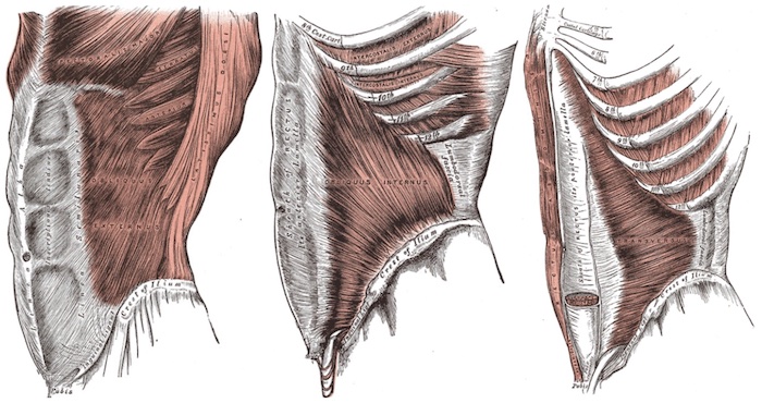
Muscles of the Lateral Abdominal Wall: External Oblique (Left), Internal Oblique (IO), and Transversus Abdominis (Right)
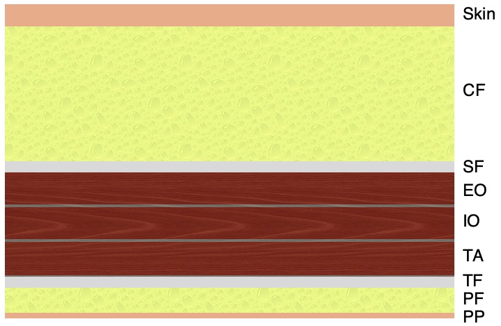
Layers of the Abdominal Wall
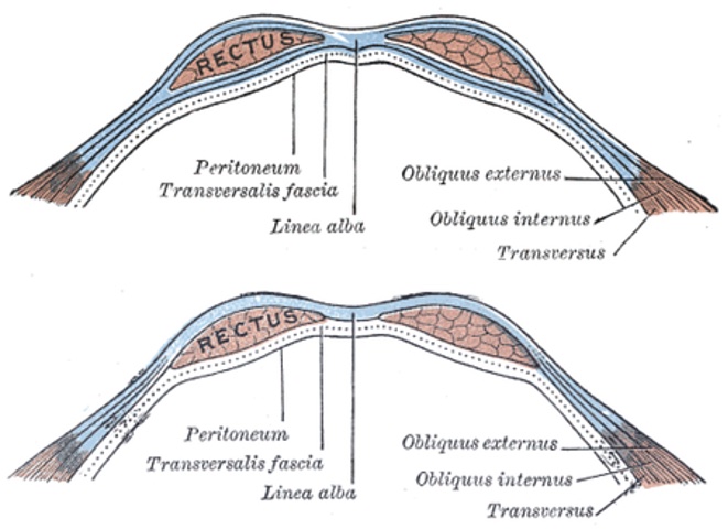
Rectus Sheath: Above (Top) and Below (Bottom) the Arcuate Line
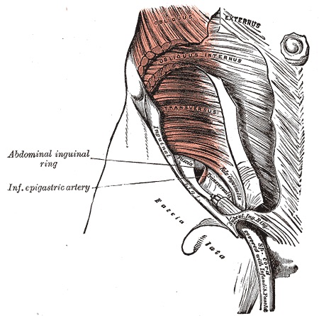
Inguinal Canal
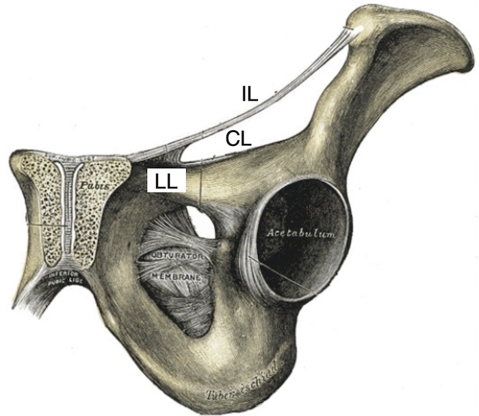
Ligaments: Inguinal (IL), Lacunar (LL), and Cooper’s (CL)
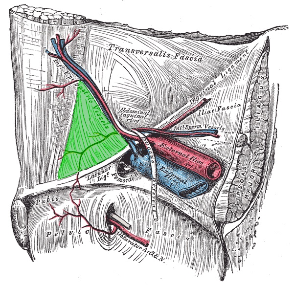
Inguinal (Hasselbach’s) Triangle
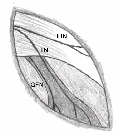
Nerves of the Inguinal Canal
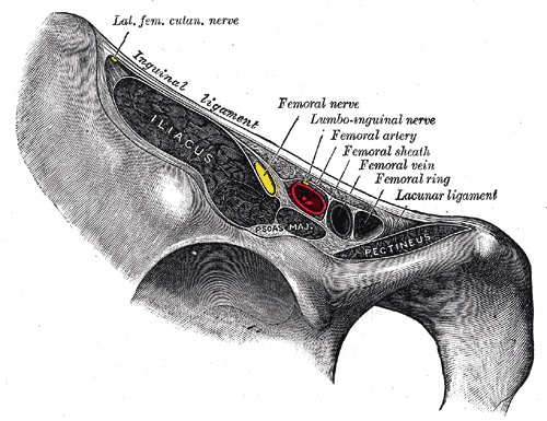
Femoral Sheath
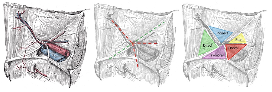
Laparoscopic Inguinal Triangles
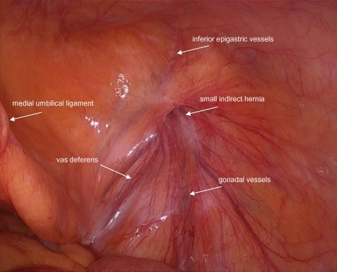
Laparoscopic View of the Inguinal Triangles 1