Adrenalectomy

Adrenalectomy

Mitchell Temple, MD

Table of Contents

Choice of Approach

Open Adrenalectomy: Reserved For Cases Unable to Be Completed Minimally Invasively, Large Tumors (> 6 cm), or Adrenal Carcinoma

Minimally Invasive Surgery (MIS) Adrenalectomy – Laparoscopic/Robotic: The Most Common Approach and Can Be Considered for Most Adrenalectomy Cases

Retroperitoneoscopic Adrenalectomy (RPA): Can Be Used Based on Surgeon Preference and in Patients with a Hostile Abdomen in Need of Adrenalectomy

Open Adrenalectomy

Patient Position

  • Supine
  • Arms Extended

Set Up

  • Incision: Ipsilateral Subcostal or Midline Incision Based on Surgeon Preference
  • Enter the Abdomen and Fully Explore to Rule Out Metastatic Disease in Adrenal Carcinoma Cases
  • Place Surgical Retractor of Preference for Optimal Exposure (i.e. Bookwalter, Thompson, Omni, etc.)

Expose the Adrenal Gland

  • Left:
    • Mobilize the Splenic Flexure Inferior and Medially
    • Mobilize the Spleen and Pancreatic Tail Medially
    • Incise the Retroperitoneum as the Spleen and Pancreas are Being Retracted and the Adrenal Gland Will Come into View
    • The Left Adrenal Vein is Exposed Working Inferiorly from the Medial Border of the Gland
      • The Left Renal Vein Can be Exposed and Used as a Landmark to Identify the Left Adrenal Vein if Needed
    • Watch for the Phrenic Vein Which May Be Seen Medially Joining the Adrenal Vein Prior to Entering the Renal Vein
  • Right:
    • Mobilize the Liver by Diving the Triangular Ligament and Lateral Attachments
    • Retract the Liver Medially to Expose the IVC and Adrenal Gland
    • Hepatic Flexure Mobilization May Be Required if Obstructing View
    • Kocher Maneuver May Be Required for Better Exposure of IVC – Usually Not Necessary
    • Expose the Right Border of the IVC to Reveal the Adrenal Vein Entering

Completion

  • Isolate the Adrenal Vein and Ligate with Suture, Clips, or Stapler
  • Adrenal Gland is Dissected Free of its Remaining Blood Supply and Attachments Using Electrocautery Until Free and Then Removed from the Field
  • Take Care Not to Rupture the Capsule to Ensure There is No Tumor Spillage
  • Ensure Hemostasis and Return Organs to Anatomical Positions
  • Close

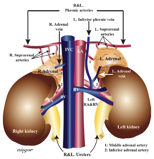
Adrenal Vasculature 1

MIS Adrenalectomy (Robotic and Laparoscopic)

Patient Position

  • Full Lateral Decubitus with Ipsilateral Side Up
  • Bed Needs to be Flexed to Open Operative Space

Port Positioning

  • Laparoscopically: 4 Ports are Placed Along the Ipsilateral Subcostal Margin (Single 10-12mm Port and Three 5mm Ports)
  • Robotically: 4 Ports are Placed Further Away from the Subcostal Margin at About a Hand’s Breadth (Need to be at Least 6 cm Apart for Appropriate Spacing to Avoid Robotic Arm Collision)
  • Additional Ports Can Be Placed as Needed for Better Retraction and/or as an Assistant Port

Set Up

  • Establish Pneumoperitoneum (Veress, OptiView, or Hasson)
  • Place Ports Under Direct Visualization
  • Dock and Target the Robot When Applicable

Expose the Adrenal Gland

  • Left:
    • Mobilize the Splenic Flexure Inferior and Medially
    • Mobilize the Spleen and Pancreatic Tail Medially
      • Use a Laparotomy Sponge under retracting instruments to avoid solid organ injury.
    • Incise the Retroperitoneum as the Spleen and Pancreas are Being Retracted and the Adrenal Gland Will Come into View
    • The Left Adrenal Vein is Exposed Working Inferiorly from the Medial Border of the Gland
      • The Left Renal Vein Can be Exposed and Used as a Landmark to Identify the Left Adrenal Vein if Needed
    • Watch for the Phrenic Vein Which May Be Seen Medially Joining the Adrenal Vein Prior to Entering the Renal Vein
  • Right:
    • Mobilize the Liver by Diving the Triangular Ligament and Lateral Attachments
    • Retract the Liver Medially to Expose the IVC and Adrenal Gland
    • Hepatic Flexure Mobilization May Be Required if Obstructing View
    • Kocher Maneuver May Be Required for Better Exposure of IVC – Usually Not Necessary
    • Expose the Right Border of the IVC to Reveal the Adrenal Vein Entering

Completion

  • Isolate the Adrenal Vein and Ligate with Clips or Stapler
  • Adrenal Gland is Dissected Free of its Remaining Blood Supply and Attachments Using Electrocautery or an Energy Device Until Free
  • Take Care Not to Rupture the Capsule to Ensure There is No Tumor Spillage
  • Remove Adrenal Gland in a Retrieval Bag
    • Depending on Tumor Size it Can Be Removed from the Larger Port, Assistant Port, or a Pfannenstiel Incision
  • Ensure Hemostasis and Return Organs to Anatomical Positions
  • Close

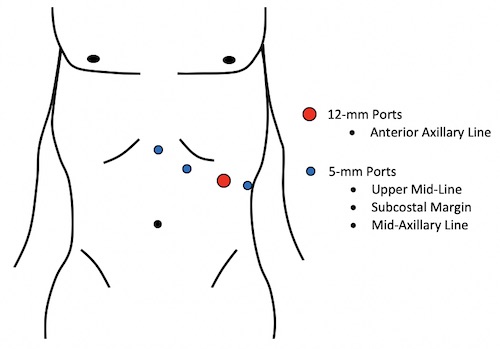
Left Laparoscopic Adrenalectomy Port Placement

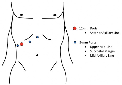
Right Laparoscopic Adrenalectomy Port Placement

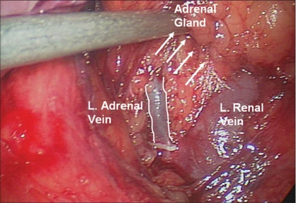
Left Adrenal Vein Draining into the Renal Vein 2

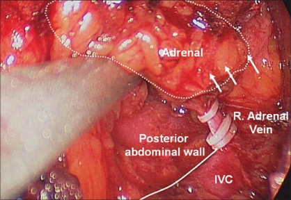
Right Adrenal Vein Draining into the IVC 2

Retroperitoneoscopic Adrenalectomy (RPA)

Patient Position

  • Prone
  • Bed Flexed to Flatten the Back and Expand the Retroperitoneal Space
  • Same for Both the Right and Left

Port Positioning

  • Generally, 3 Ports are Placed in a Horizontal Line 4-5 cm Inferior to Inferior Edge of the 12th Rib
    • Central 10 mm Balloon Trocar
    • One 5 mm Port Laterally
    • One 5mm Port Medially
  • Additional Ports Can Be Placed as Needed for Better Retraction and/or as an Assistant Port

Set Up

  • Insert the Central Balloon 10 mm Trocar via Open Cutdown
  • Place 5 mm Trocars Laterally and Medially, Guided Manually Through the Central Incision
  • Establish Insufflation (18-20 mmHg)

Dissection

  • Perirenal Fat is Dissected Lateral-to-Medial to Define the Upper Pole of the Kidney and Separate from the Inferior Border of the Adrenal Gland
  • Adrenal Gland is Dissected Using Blunt and Sharp Dissection to the Paraspinous Muscles Medially and the Peritoneum Laterally
  • Leave the Superior Attachment so That the Adrenal Gland Remains Suspended Until the End of the Operation
  • Medially Dissect Off the Renal Hilum by Diving the Attachments Halfway Up the Adrenal Gland
  • Expose the Adrenal Vein:
    • Left: Medial/Inferior Dissection Exposes the Adrenal Vein – Phrenic Vein May Be Seen Medially Joining the Adrenal Vein Prior to Entering the Renal Vein and Can Be Divided as Needed
    • Right: Medial Dissection Exposes the IVC and Adrenal Vein

Completion

  • Isolate the Adrenal Vein and Ligate with Clips
  • Retract the Gland by the Remaining Vein to Dissect the Final Superior Attachments
    • Use a Vessel Sealing Device to Ligate the Adrenal Arterial Supply as Encountered
  • Take Care Not to Rupture the Capsule to Ensure There is No Tumor Spillage
  • Remove Adrenal Gland in a Retrieval Bag
  • Ensure Hemostasis
  • Close

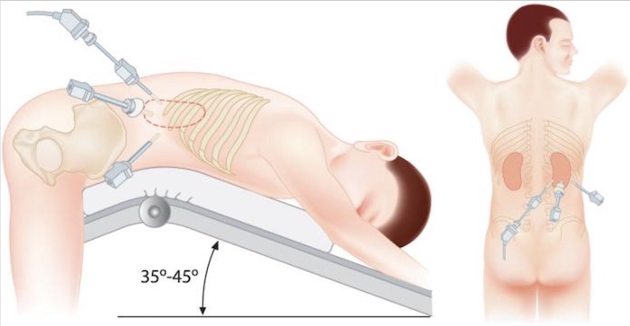
Retroperitoneoscopic Port Placement and Patient Positioning for Right Adrenalectomy 3

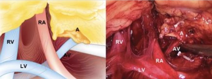
Left Retroperitoneoscopic View: Adrenal (A), Adrenal Vein (AV), Lumbar Vein (LV), Renal Vein (RV), and Renal Artery (RA) – Note Clips for the Inferior and Medial Adrenal Arteries 3

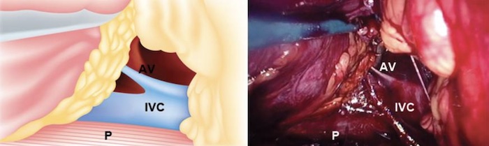
Right Retroperitoneoscopic View: Adrenal Vein (AV), IVC, and Psoas Muscle (P) 3

Complications

Bleeding/Retroperitoneal Hematoma

Surgical Site Infection (SSI)

Adrenal insufficiency

Iatrogenic Injury

  • Right: IVC or Liver Injury
  • Left: Spleen, Colon, or Pancreas Injury

References

Cover: Raffaelli M, De Crea C, Bellantone R. Laparoscopic adrenalectomy. Gland Surg. 2019 Jul;8(Suppl 1):S41-S52. (License: CC BY-NC-4.0)

  1. Uludağ M, Aygün N, İşgör A. Surgical Indications and Techniques for Adrenalectomy. Sisli Etfal Hastan Tip Bul. 2020 Mar 24;54(1):8-22. (License: CC BY-NC-4.0)
  2. Mellon MJ, Sethi A, Sundaram CP. Laparoscopic adrenalectomy: Surgical techniques. Indian J Urol. 2008 Oct;24(4):583-9. (License: CC Not Specified)
  3. Hisano M, Vicentini FC, Srougi M. Retroperitoneoscopic adrenalectomy in pheochromocytoma. Clinics. 2012;67:161-167. (License: CC BY-NC 3.0)
  4. Sippel R, Chen H. The Handbook of Endocrine Surgery . World Scientific; 2012.
  5. Dimick JB, Upchurch GR, Sonnenday CJ, Kao LS. Clinical Scenarios in Surgery : Decision Making and Operative Technique. Wolters Kluwer; 2019.