Breast: Anatomy & Physiology
Anatomy
Structure
- External Features:
- Nipple – Raised Tissue Through Which Milk is Secreted
- Areola – Pigmented Region Around the Nipple
- Montgomery’s Glands – Modified Sweat Glands in the Areola
- Oily Secretions Protect Nipple During Breastfeeding
- Montgomery’s Glands – Modified Sweat Glands in the Areola
- Internal Features:
- Cooper’s Ligaments (Suspensory Ligaments of Cooper) – Fibrous Ligaments Radiate from the Superficial Fascia to the Skin to Maintain Structural Integrity
- Tail of Spence (Axillary Process) – Extension of Breast Tissue into the Axilla
- Mammary Glands:
- Lobules – Groups of Alveoli Which Produce Milk
- Lactiferous (Milk/Galactophorous) Ducts – Drain Milk from the Lobule to the Nipple
- Each Breast Has 15-20 Ducts
- Diameter: 2.0-4.5 mm
- The Upper Outer Quadrant (UOQ) Contains the Majority of the Epithelial Tissue
- Explains Why the UOQ is the Most Common Site of Both Benign & Malignant Breast Disease – Most are Derived from Epithelial Tissue
Described Quadrants
- Upper Outer Quadrant (UOQ)
- Upper Inner Quadrant (UIQ)
- Lower Outer Quadrant (LOQ)
- Lower Inner Quadrant (LIQ)
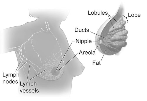
Breast Anatomy 1
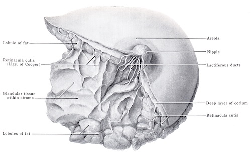
Cooper’s Ligaments 2
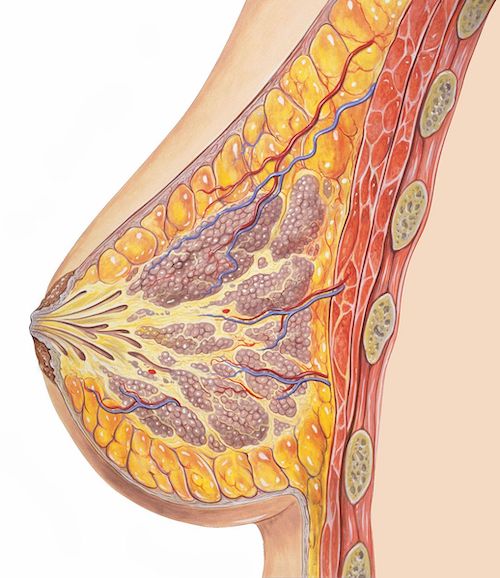
Breast Cross-Section 3
Density
- Fibrous & Glandular Tissue Increase Density
- Fatty Tissue Decreases Density
- Breasts with Increased Density Increase the Difficulty of Identifying Pathologic Lesions on Mammogram
- BI-RADS Classification:
- Class A: Almost Entirely Fatty (10%)
- Class B: Scattered Fibroglandular Density (40%)
- Class C: Heterogeneously Dense (40%)
- Class D: Extremely Dense (10%)
- *Class C & D are Considered Dense Breasts
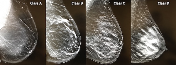
Breast Density
Vascular Supply
- Medial Breast
- Internal Mammary/Thoracic Artery (IMA) (From Subclavian)
- Lateral Breast
- Lateral Thoracic Artery (From Axillary)
- Thoraco-Acromial Artery (From Axillary)
- Intercostal Arteries
- Batson’s Plexus Mn
- Valveless Veins of Paravertebral Space
- Connected to Thorax (Breast) and Deep Pelvic Veins (Rectum/Prostate)
- Reason for Mets to Spine (Often First)
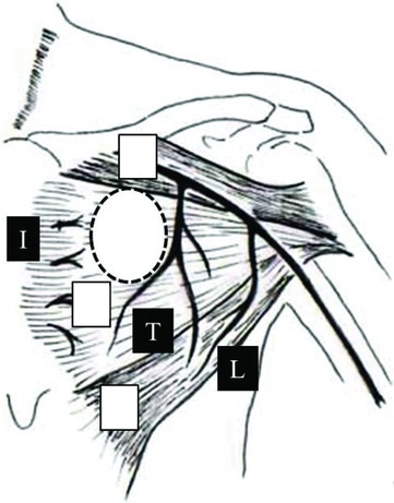
Breast Blood Supply: Internal Mammary Artery (I), Thoracoacromial Artery (T), Lateral Thoracic Artery (L) 4
Nerves
- Long Thoracic Nerve
- Location: Medial Border of Axilla Along Chest Wall
- Innervate: Serratus Anterior
- Deficit: Winged Scapula
- Thoracodorsal Nerve
- Location: Mid-Axilla Just Below Axillary Vein
- Innervate: Latissimus Dorsi
- Deficit: Weak Arm Adduction (Pull Ups) & Internal Rotation
- Medial Pectoral Nerve Mn
- Innervate: Pectoralis Major & Minor
- Lateral Pectoral Nerve
- Innervate: Pectoralis Major
- Deficit: Muscle Atrophy & Limited Shoulder Motion
- Intercostobrachial Nerve
- The Lateral Cutaneous Branch of the Second Intercostal Nerve
- Innervate: Sensation to Medial Arm/Axilla
- Deficit: Loss of Sensation to Medial Arm
- Most Common Injured Nerve in MRM/ALND
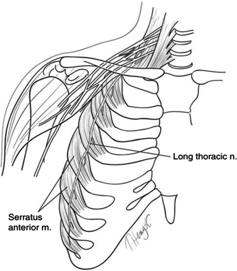
Long Thoracic Nerve 5

Winged Scapula 6
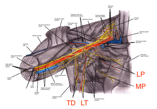
Axillary Nerves 7
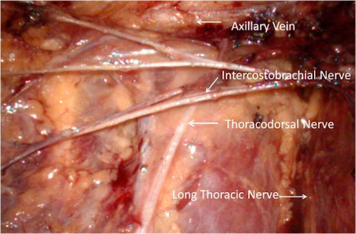
Intercostal Brachial Nerve 8
Lymphatics
- Initial Lymph Nodes
- Pectoral: Lateral
- Interpectoral Nodes: Superior
- Parasternal Nodes: Medial/Superior
- Subphrenic Nodes: Medial/Inferior (Liver/Abd Wall Mets)
- Anastomoses: Contralateral Breast
- Sappey Plexus Mn
- Subareolar Lymphatic Plexus
- Where All Lymphatics of the Breast Converge
- Place for Injection During SLNB
- Drainage
- Axillary Lymph Nodes (97%)
- Level I: Lateral to Pectoralis Minor
- Level II: Deep to Pectoralis Minor
- Rotter’s Nodes: Between Pectoralis Minor & Major
- Level III: Medial to Pectoralis Minor
- Internal Mammary Nodes (2%)
- Axillary Lymph Nodes (97%)
- Final Drainage
- Right Lymphatic Duct: Upper Right Side
- Thoracic Duct: Everything Else
- Supraclavicular Lymph Nodes
- Final Nodes at Vascular Emptying
- Left (Virchow’s Nodes)
- Sign of Intraabdominal Malignancy
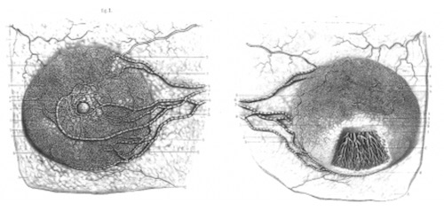
Sappey Plexus 9
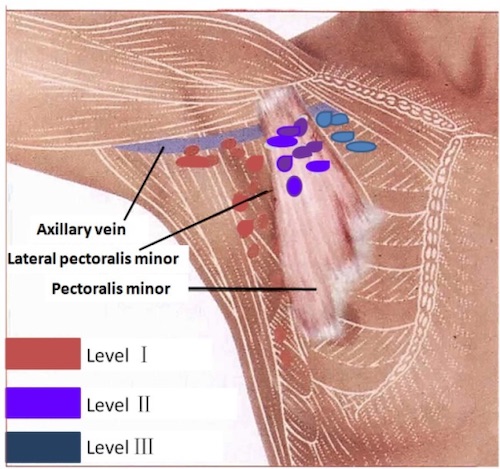
Axillary Lymph Nodes Levels 10
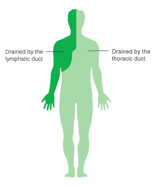
Body Lymphatic Drainage 11
Physiology
Hormones
- Estrogen Mn
- Duct Development
- Glandular Growth
- Breast Swelling
- Progesterone
- Lobule Development
- Glandular Maturation
Mnemonics
Batson vs Sappey Plexus
- B-B: Batson – Back
- S-S: Sappey – Subareolar
Pectoral Nerve Innervation
- M-M: Medial Gets Both M’s (Major & Minor)
- L-L: Lateral Only Gets Large (Major)
Estrogen vs Progesterone Effects on Breast
- E-E: Double-E Grows in Size
- Pro-Pro: Professionals are Mature
References
- National Cancer Institute. Wikimedia Commons. (License: Public Domain)
- Grant JCB. Wikimedia Commons. (License: Public Domain)
- Meyerson A. Wikimedia Commons. (License: CC BY-SA-3.0)
- Asamura S, Kurita T, Motoki K, Yasuoka R, Hashimoto T, Isogai N. Efficacy and feasibility of the submuscular implantation technique for an implantable cardiac electrical device. Eplasty. 2014 Oct 30;14:e40. (License: CC BY-2.0)
- Martin RM, Fish DE. Scapular winging: anatomical review, diagnosis, and treatments. Curr Rev Musculoskelet Med. 2008 Mar;1(1):1-11. (License: CC BY-NC-2.0)
- Wikimedia Commons. (License: CC BY-SA-3.0)
- Chandler MH, Dimatteo L, Hasenboehler EA, Temple M. Intraoperative brachial plexus injury during emergence following movement with arms restrained: a preventable complication? Patient Saf Surg. 2007 Dec 19;1:8. (License: CC BY-2.0)
- Du J, Liang Q, Qi X, Ming J, Liu J, Zhong L, Fan L, Jiang J. Endoscopic nipple sparing mastectomy with immediate implant-based reconstruction versus breast conserving surgery: a long-term study. Sci Rep. 2017 Mar 31;7:45636. (License: CC BY-4.0)
- Suami H, Pan WR, Mann GB, Taylor GI. The lymphatic anatomy of the breast and its implications for sentinel lymph node biopsy: a human cadaver study. Ann Surg Oncol. 2008 Mar;15(3):863-71. (License: CC BY-NC-2.0)
- Lu Q, Hua J, Kassir MM, Delproposto Z, Dai Y, Sun J, Haacke M, Hu J. Imaging lymphatic system in breast cancer patients with magnetic resonance lymphangiography. PLoS One. 2013 Jul 5;8(7):e69701. (License: CC Not Specified)
- Cancer Research UK. Wikimedia Commons. (License: CC BY-SA-4.0)