Esophagus: Anatomy & Physiology
Anatomy
Layers
- Mucosa (Squamous Epithelium)
- Submucosa
- Muscularis Propria (Longitudinal)
- Upper 1/3: Striated
- Lower 2/3: Smooth
- No Serosa
- Importance: CA Spreads Through Lymphatics
Sphincters
- Upper Esophageal Sphincter (UES)
- Cricopharyngeus Muscle
- 15 cm From Incisors
- Most Common Site of Iatrogenic Perforation and Foreign Body
- Prevents Air Swallowing
- Innervation: Recurrent Laryngeal Nerve
- Contracted at Rest
- Failure to Relax After CVA Causes a Risk for Aspiration
- Lower Esophageal Sphincter (LES)
- Not True Anatomic Sphincter – From Inner Circular Muscle Layer
- 40 cm From Incisors
- Prevents Reflux
- Innervation: Vagus
- High Resting Tone, Relaxes with Swallowing or Gastric Distention
Structure
- Killian Triangle
- Boundaries:
- Superior/Lateral: Inferior Constrictors
- Inferior: Cricopharyngeus
- Site of Zenker’s Diverticulum
- Boundaries:
- Anatomic Sites of Narrowing:
- Upper Esophageal Sphincter (UES) – Most Narrow
- Left Mainstem Bronchus
- Aortic Arch
- Diaphragm
Blood Supply
- Arterial Supply:
- Cervical Esophagus: Inferior Thyroid Artery
- Thoracic Esophagus: Bronchial Arteries & Vessels Directly from Aorta
- Abdominal Esophagus: Left Gastric Artery & Inferior Phrenic Artery
- Venous Drainage:
- Azygos Vein
- Hemi-Azygos Vein
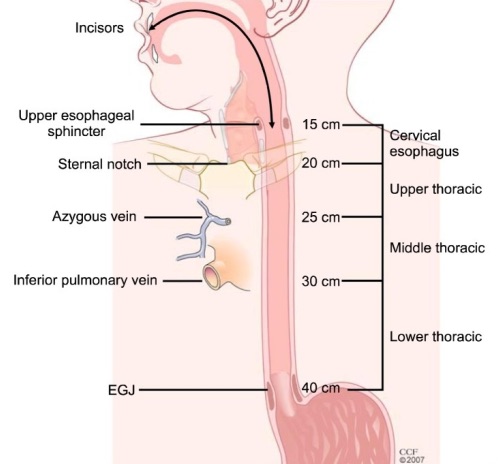
General Esophagus Distances 1
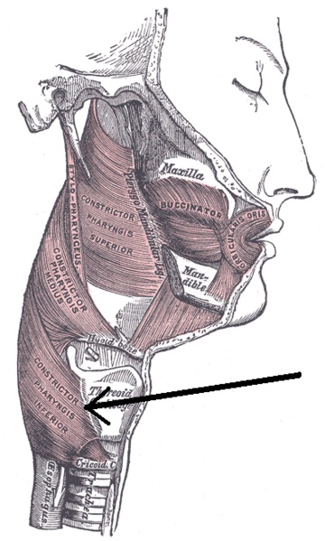
UES 2
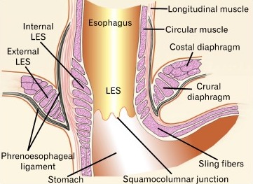
LES 3
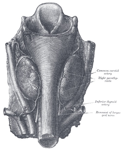
Blood Supply of the Cervical Esophagus 2
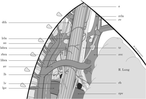
Blood Supply of the Thoracic Esophagus 4
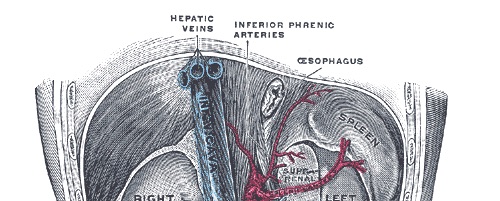
Blood Supply of the Abdominal Esophagus 2
Lymphatics
- Upper 2/3: Superior
- Cervical Esophagus – Deep Cervical LN
- Thoracic Esophagus – Paraesophageal & Mediastinal LN
- Lower 1/3: Inferior
- Abdominal Esophagus – Gastric/Cardiac & Celiac LN
- Drain into Cisterna Chyli/Thoracic Duct
Nerves
- Right Vagus
- Twists Over the Posterior Aspect
- *Twists Like A Screw Going Down (Clockwise)
- Innervates the Celiac Plexus
- Twists Over the Posterior Aspect
- Left Vagus
- Twists Over the Anterior Aspect
- Innervates the Liver & Biliary Tree
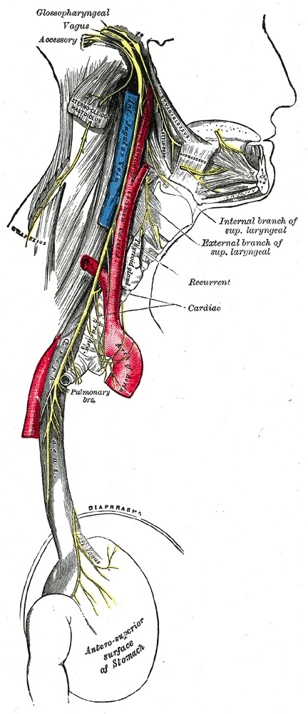
Vagus Nerve 2
Physiology
Swallowing Stages
- 1. Oral Phase: Mastication, Trough Formation & Bolus Movement Posteriorly
- 2. Pharyngeal Phase: Close Oropharynx/Larynx & Bolus Passes Pharynx
- 3. Peristalsis & Relaxation
Peristalsis
- Propagation of Food Bolus by Contraction of Circular Muscles in Wave Formation
- Occludes Proximally and Propels Food Distally
- Types:
- Primary Peristalsis:
- Initiated by Food Bolus & Swallowing
- Normal Peristaltic Contraction
- Secondary Peristalsis:
- Initiated by Distention (Retained Bolus/Reflux)
- Propagates Distally
- Tertiary Contraction:
- Dysfunctional Contractions
- Does Not Propagate/Peristalse
- Primary Peristalsis:
Normal Manometry Pressures
- Upper Esophageal Sphincter (UES)
- Rest: 50-70 mmHg
- Swallowing: 15 mmHg
- Lower Esophageal Sphincter (LES)
- Rest: 10-20 mmHg
- Swallowing: 0 mmHg
References
- Rice TW. Esophageal Cancer Staging. Korean J Thorac Cardiovasc Surg. 2015 Jun;48(3):157-63. (License: CC BY-NC-3.0)
- Gray H. Anatomy of the Human Body (1918). Public Domain.
- Mittal RK, Hong SJ, Bhargava V. Longitudinal muscle dysfunction in achalasia esophagus and its relevance. J Neurogastroenterol Motil. 2013 Apr;19(2):126-36. (License: CC BY-NC-3.0)
- Cuesta MA, van der Wielen N, Weijs TJ, Bleys RL, Gisbertz SS, van Duijvendijk P, van Hillegersberg R, Ruurda JP, van Berge Henegouwen MI, Straatman J, Osugi H, van der Peet DL. Surgical anatomy of the supracarinal esophagus based on a minimally invasive approach: vascular and nervous anatomy and technical steps to resection and lymphadenectomy. Surg Endosc. 2017 Apr;31(4):1863-1870. (License: CC BY-4.0)