
Esophageal Motility Disorders
Esophageal Motility Disorders
Yaiel G. Rodríguez-Avilés, MD
Table of Contents
Definition
Definition: A Disorder of Esophageal Motor Function Characterized by Abnormal Esophageal Peristalsis, Abnormal Esophagogastric Junction (EGJ) Outflow, or Impaired Lower Esophageal (LES) Relaxation with Resulting Impairment of Esophageal Bolus Transit
Also Known as Esophageal Dysmotility
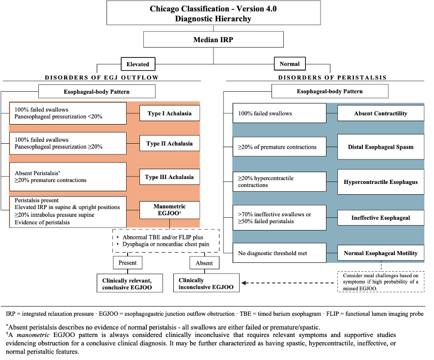
Chicago Classification – Version 4.0 (CCv4)
- Standardized Diagnostic System That Categorizes Esophageal Motility Disorders Based on High-Resolution Manometry (HRM) Findings to Guide Clinical Assessment and Treatment
- The Classification is Hierarchical and Divides Essentially Separates Disorders into Two Groups:
- Disorders of EGJ Outflow
- Disorders of Peristalsis
Key Changes from CCv3
- Incorporates Both Supine and Upright Swallows
- Adds Provocative Maneuvers, Including Multiple Rapid Swallows and the Rapid Drink Challenge
- Eliminates the Distinction Between “Major” and “Minor” Disorders of Peristalsis
- Removes Fragmented Peristalsis as a Standalone Diagnosis and Incorporates Fragmented Contractions into the Assessment of Ineffective Esophageal Motility
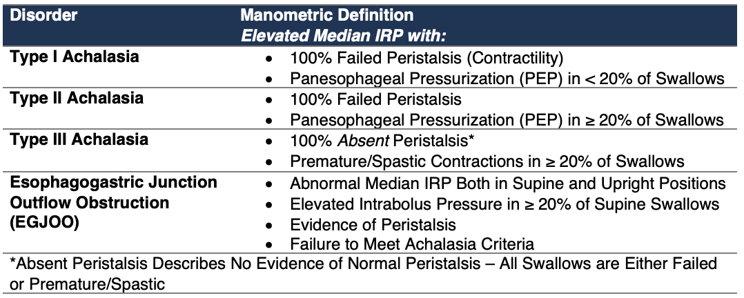
- Clarifies the Definition of Type III Achalasia, Which Requires 100% Absent Normal Peristalsis with Premature (Spastic) Contractions
- Makes the Diagnostic Criteria for EGJOO More Stringent
- Emphasizes That EGJOO, Distal Esophageal Spasm, and Hypercontractile Esophagus Cannot Be Considered Clinically Relevant Based on Manometry Alone
- Raises the Threshold for a Conclusive Diagnosis of Ineffective Esophageal Motility
Classification/Disorders
Disorders of EGJ Outflow
Disorders of Peristalsis

Diagnostic Evaluation
Clinical Presentation
- Dysphagia – The Most Common Symptom
- Chest Pain
- Regurgitation
- Heartburn – Can Overlap with Symptoms of Gastroesophageal Reflux Disease (GERD)
- Globus Sensation
- Food Retention
- Cough
- Recurrent Aspiration
- Halitosis
- Weight Loss
- Symptoms are Generally Nonspecific
Diagnostic Evaluation
- Initial Evaluation: Assess Symptoms and Exclude Mechanical Obstruction with Upper Endoscopy
- High-Resolution Manometry (HRM): Primary Diagnostic Test for Characterizing Esophageal Motility and Classifying Motility Disorders According to CCv4
- Supportive Testing: Timed Barium Esophagram (TBE) and/or Functional Lumen Imaging Probe (FLIP) May Provide Additional Evidence of EGJ Outflow Obstruction or Impaired Esophageal Emptying
- Clinical Correlation: Manometric Findings Should Be Interpreted in the Context of Symptoms and Supportive Testing, Particularly for EGJOO and Other Potentially Clinically Inconclusive Motility Patterns
Indications for HRM
- Esophageal Dysphagia
- Gastroesophageal Reflux Disease (GERD) Resistant to Standard Therapy
- Evaluation Before Antireflux Surgery to Exclude Motility Disorder that May Alter Surgical Planning (Full vs Partial Fundoplication)
- Non-Cardiac Chest Pain
- Suspected Systemic Diseases Affecting Smooth Muscle or the Autonomic Nervous System (e.g. Scleroderma, Collagen Vascular Diseases)
Limitations of HRM
- HRM is Not Diagnostic for GERD and Does Not Determine its Severity
- HRM is Not the Primary Test for Oropharyngeal Dysphagia
- Modified Barium Swallow or Video-Fluoroscopy is Generally Preferred
- Prior Foregut Surgery or Invasive EGJ Treatment (e.g., Botox, Bariatric Surgery) Can Alter Normal Anatomy and Limit Interpretation
Supportive Testing
- Upper Endoscopy: Excludes Structural or Mucosal Disease and Pseudoachalasia; Mechanical Obstruction Changes Interpretation
- Timed Barium Esophagram (TBE) with Tablet: Assesses Objective Esophageal Emptying and Supports EGJ Obstruction
- Functional Lumen Imaging Probe (FLIP): Measures EGJ Distensibility and Contractile Response; Particularly Useful for Equivocal Achalasia or EGJOO
- HRM with Impedance: Adds Information About Bolus Transit and Clearance
How to Interpret High-Resolution Manometry (HRM)
Basics
- HRM Uses a Catheter Containing Closely Spaced Pressure Sensors to Simultaneously Measure Pressure from the Pharynx Through the Esophagus and Into the Stomach
- The Results Are Displayed as Esophageal Pressure Topography
- Horizontal Axis: Time
- Vertical Axis: Anatomic Position
- Cool Colors: Lower Pressure
- Warm Colors: Higher Pressure
HRM Assesses
- Esophagogastric Junction (EGJ) and Lower Esophageal Sphincter (LES) Relaxation
- Esophageal Body Peristalsis
- Contraction Timing and Vigor
- Esophageal Pressurization
- Bolus Transit When Impedance is Incorporated
HRM Metrics
- Integrated Relaxation Pressure (IRP)
- Distal Contractile Integral (DCI)
- Distal Latency (DL)
- Contractile Wavefront Integrity
- Pressurization
Integrated Relaxation Pressure (IRP)
- Measures the Mean Nadir Pressure of the EGJ During the 4 Seconds of Greatest EGJ Relaxation Within a 10-Second Window Following Swallow Initiation
- Describes How Effectively the EGJ Relaxes During Swallowing
- Diagnostic Threshold:
- Abnormal/Elevated Thresholds Depend on the Catheter System and Patient Position
- Medtronic Systems
- Supine: ≥ 15 mmHg
- Upright: ≥ 12 mmHg
- Laborie/Diversatek Systems
- Supine: ≥ 22 mmHg
- Upright: ≥ 15 mmHg
- A Common Teaching/ABSITE Cutoff is Usually ≥ 15 mmHg
Distal Contractile Integral (DCI)
- Measures the Vigor of Distal Esophageal Contraction by Integrating:
- Amplitude (mmHg)
- Duration (s)
- Length (cm)
- Diagnostic Threshold:
- Hypercontractile: > 8,000 mmHg·s·cm
- Normal: 450-8000 mmHg·s·cm
- Weak: 100-449 mmHg·s·cm
- Failed: < 100 mmHg·s·cm
Contractile Wavefront Integrity
- Describes the Continuity of Peristaltic Contractions
- Diagnostic Threshold:
- Fragmented Swallow: Normal DCI (≥ 450 mmHg·s·cm) with a Large Transition Zone Defect/Break of Peristalsis (> 5 cm) Under a 20-mmHg Isobaric Contour
- Fragmented Peristalsis is No Longer a Standalone Diagnosis in CCv4 and Is Incorporated Into the Assessment of IEM
Distal Latency (DL)
- Contractile Deceleration Point (CDP): The Inflection Point at Which the Peristaltic Contraction Transitions from Its Rapid Proximal Phase to Its Slower Distal Phase (Often About 3 cm Proximal to the LES)
- Physiologically, This Likely Represents Conversion from Smooth Muscle Esophageal Body Contraction to the LES Contraction
- Distal Latency (DL): Measures the Interval from Swallow Initiation (Upper Esophageal Sphincter Relaxation) to the Contractile Deceleration Point (CDP)
- Diagnostic Threshold:
- Premature/Spastic Contraction: DL < 4.5 Seconds with DCI ≥ 450 mmHg·s·cm
Pressurization
- Represents Increased Pressure When a Bolus or Gas Pocket Becomes Entrapped Between Adjacent Contracting Muscle Segments or is Obstructed by a Non-Relaxing Valve
- Diagnostic Threshold:
- Panesophageal Pressurization: Pressure ≥ 30 mmHg Extending from the Upper Esophageal Sphincter (UES) to the EGJ
- Intrabolus Pressurization: Localized Pressure Generated Proximal to an Obstructed EGJ; ≥ 20 mmHg in the Supine Position Supports EGJ Outflow Obstruction on Medtronic Systems
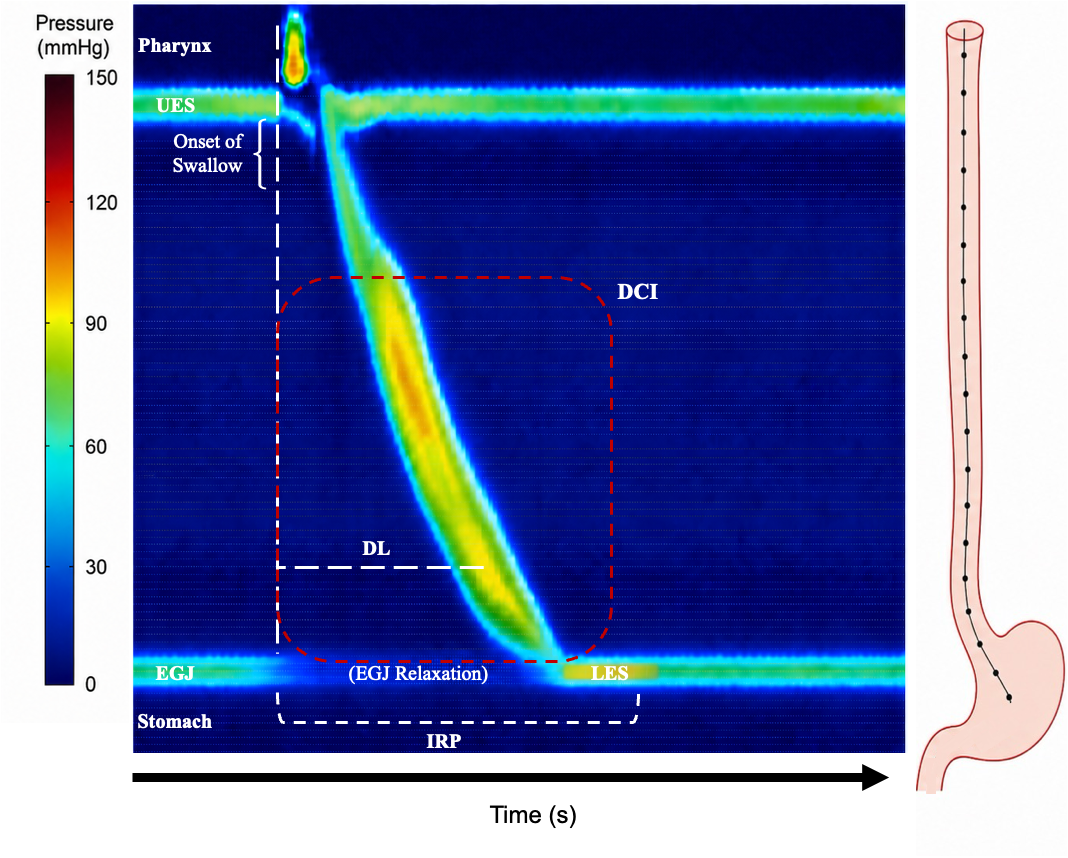
High-Resolution Manometry with Esophageal Pressure Topography: Swallow Initiation is Marked by Pharyngeal Contraction and UES Relaxation, Followed by an Intact, Sequentially Propagated Esophageal Contraction and Coordinated EGJ/LES Relaxation. DCI Reflects the Vigor of the Distal Esophageal Contraction, DL Measures the Interval From UES Relaxation to the CDP, and IRP Represents EGJ Relaxation During the Post-Swallow Period. Pressure Magnitude Is Indicated by the Color Gradient.