Trauma: Hand Injury
Hand Injury
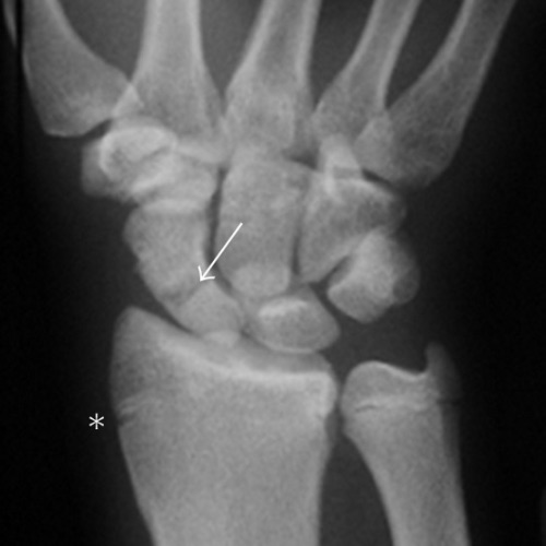
Scaphoid Fracture
- The Most Common Carpal Bone Fracture
- Complications:
- High Rate of Nonunion (5-25%)
- Avascular Necrosis (5-10%)
- Presentation: “Snuffbox” Tenderness
- Immediate X-Ray May Be Negative
- CT – Best Modality for Nonunion
- MRI – If Concerned for Osteonecrosis
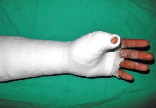
- Treatment: Thumb Spica Cast
- May Consider Operative Repair (ORIF) if Significantly Displaced or Unstable

Scaphoid Fracture 1
![]()
Thumb Spica Cast 2
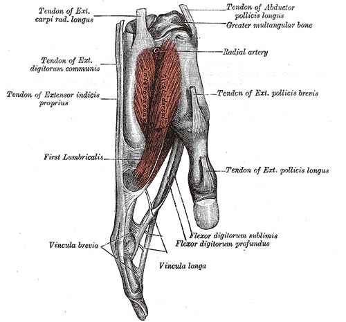
Flexor Tendons
- Tendons:
- Flexor Digitorum Superficialis (FDS): To Proximal Interphalangeal (PIP) Joint
- Flexor Digitorum Profundus (FDP): To Distal Interphalangeal (DIP) Joint
- Flexor Pollicis Longus (FPL): To the Thumb
- Flexor Carpi Radialis (FCR): Primary Wrist Flexor
- Flexor Carpi Ulnaris (FCU): Primary Wrist Flexor
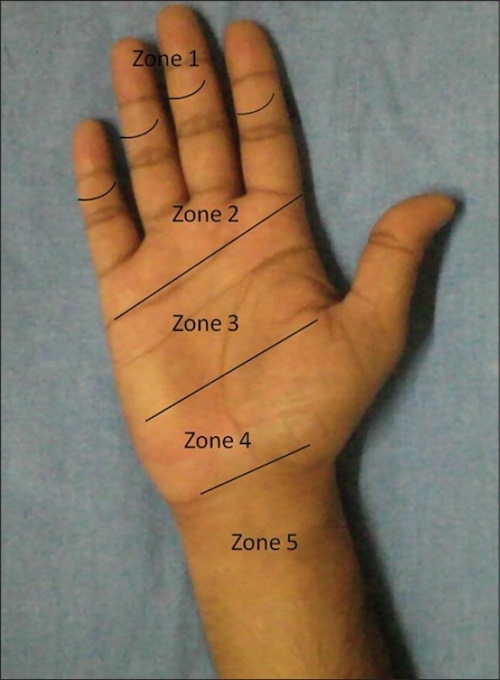
- Flexor Zones:
- Zone I: Distal to the FDS Insertion
- Zone II: FDS Insertion to the Distal Palmar Crease (Proximal A1 Pulley)
- Zone III: Palm
- Zone IV: Carpal Tunnel
- Zone V: Forearm to the Carpal Tunnel
- Injuries:
- Jersey Finger: Avulsion of the FDP
- Finger Sits in Slight Extension
- Jersey Finger: Avulsion of the FDP
- Primarily Repair Tendon Lacerations > 50-60% Width

Finger Flexor Tendons 3

Finger Flexor Zones 4
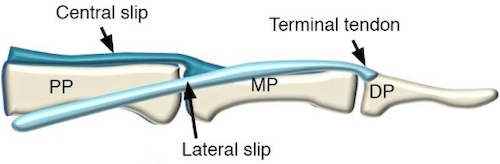
Extensor Tendons
- Tendons:
- Central Slip: To Proximal Interphalangeal (PIP) Joint
- Lateral Bands: To Distal Interphalangeal (DIP) Joint
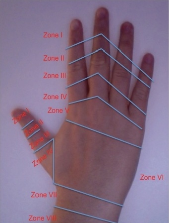
- Extensor Zones:
- Zone I: Distal to DIP Joint (Or IP of the Thumb)
- Zone II: Middle Phalanx (Or Proximal Phalanx of the Thumb)
- Zone III: PIP Joint (Or MCP Joint of the Thumb)
- Zone IV: Proximal Phalanx (Or Metacarpal of the Thumb)
- Zone V: MCP Joint (Or CMC of the Thumb)
- Zone VI: Disruption Over the Metacarpal
- Zone VII: Wrist Joint
- Zone VIII: Distal Forearm
- Injuries:
- Mallet Finger: Avulsion of Terminal Extensor Tendon Over DIP
- Characterized by DIP Flexion
- Boutonniere Deformity: Rupture of Central Slip Over PIP
- Characterized by PIP Flexion & DIP Extension
- Mallet Finger: Avulsion of Terminal Extensor Tendon Over DIP
- Primarily Repair Tendon Lacerations > 50-60% Width

Finger Extensor Zones 5

Finger Extensor Tendons 6
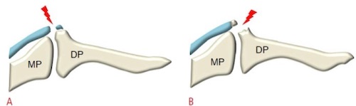
Mallet Finger 6
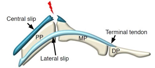
Boutonniere Deformity 6
Thumb Injury
- Base of Thumb Fracture
- Types:
- Extra-Articular
- Partial Intra-articular (Bennett)
- Complete Intra-articular (Rolando)
- Often Caused by an Axial Force Applied to the Thumb in Flexion
- Image with True AP Thumb X-Rays
- Consider CT if Fracture Appears Complex
- Primary Treatment: Closed Reduction and Thumb Spica Cast
- May Consider ORIF or Percutaneous K-Wire in Select Scenarios
- Types:
- Collateral Ligament Injury
- Ulnar and Radial Collateral Ligaments
- UCL Injury is 10x More Common
- Radially Directed Force Caused Hyper-Abduction of the MCP Joint
- Causes Pain with Pinch or Grasp
- Imaging: Radiograph (Can Do Stressed Views)
- UCL Injury – Avulsion or Condylar Fracture
- RCL Injury – Pronation of Proximal Phalanx
- MRI or Ultrasound are Available as Well
- Primary Treatment: Immobilization with Thumb Spica Cast
- May Consider Operative RCL/UCL Repair in Select Scenarios
- Ulnar and Radial Collateral Ligaments
Digit Replantation Indications
- All Thumb Injuries
- Multiple Digits
- Zone I (DIP & Distal)
- Zone 2 (MCP & PIP) Have Poor Outcomes
- Pediatrics
References
- Nakamoto JC, Saito M, Medina G, Schor B. Scaphoid stress fracture in high-level gymnast: a case report. Case Rep Orthop. 2011;2011:492407. (License: CC BY-3.0)
- Sasi PK, Mahapatra S, Raj Pallapati SC, Thomas BP. Acute Traumatic Musculotendinous Avulsion of the Flexor Pollicis Longus Tendon Treated with Primary Flexor Digitorum Superficialis Transfer: A Novel Technique of Management. Case Rep Orthop. 2016;2016:2106203. (License: CC BY-4.0)
- Gray H. Anatomy of the Human Body (1918). Public Domain.
- Kotwal PP, Ansari MT. Zone 2 flexor tendon injuries: Venturing into the no man’s land. Indian J Orthop. 2012 Nov;46(6):608-15. (License: CC BY-NC-SA-3.0)
- Griffin M, Hindocha S, Jordan D, Saleh M, Khan W. Management of extensor tendon injuries. Open Orthop J. 2012;6:36-42. (License: CC BY-NC-3.0)
- Lee SA, Kim BH, Kim SJ, Kim JN, Park SY, Choi K. Current status of ultrasonography of the finger. Ultrasonography. 2016 Apr;35(2):110-23. (License: CC BY-NC-3.0)