Intraoperative Cholangiogram (IOC)
David Ray Velez, MD
The Operative Review of Surgery. 2023; 1:48-51.
Table of Contents
Indications
Definition
- Intraoperative Fluoroscopic X-Ray Imaging with Contrast Through the Bile Ducts
- Goals: 1
- Identify Bile Duct Stones
- Clarify Biliary Anatomy
- Prevent Bile Duct Injuries
Indications 2
- Choledocholithiasis Diagnosed Preoperatively
- Concern for Possible Choledocholithiasis
- Jaundice
- Gallstone Pancreatitis
- Elevated Liver Function Tests
- Dilated Common Bile Duct > 5-7 mm
- Dilated Cystic Duct > 3 mm
- Multiple Small Stones in the Gallbladder
- Presumed Choledocholithiasis that Passed (Marginal Improvement in Labs)
- Need to Delineate Unclear Ductal Anatomy
- Concern for Possible Bile Duct Injury or Leak
- Allows Earlier Recognition of Injury
- Prevents Complete CBD Transection (Does Not Prevent Injury)
Routine vs Selective Use 3,4
- Routine Use is Controversial – Evidence is Insufficient
- Currently Considered Not Mandatory, Although Practice May Improve Outcomes in More Challenging Cases
- Should Be Used Liberally Regardless
- Likelihood of Finding an Unsuspected Stone: 3-7% 5

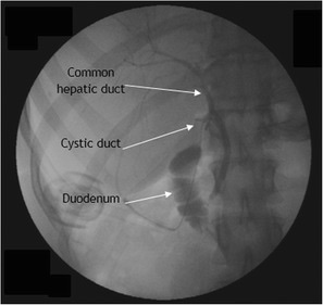
Appropriate Visualization on IOC 7
Technique
Clamps
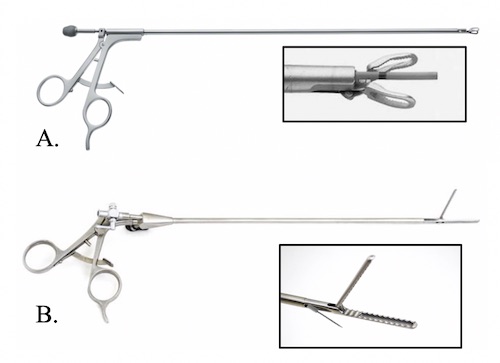
- Olsen Clamp – Clamp onto the Cystic Duct with a Blunt Catheter Extending Out of the Center of the Clamp
- Kumar Clamp – Clamp onto the Infundibulum with a Sharp 19 ga Needle Extending Out of the Side of the Clamp
- *No Evidence to Suggest that Any Technique is Superior to the Another
Olsen Clamp Technique
- Obtain the Critical View of Safety as Normal
- Place a Clip Proximally Across the Junction of the Gallbladder Infundibulum & Cystic Duct
- Prevents Reflux of Contrast into the Gallbladder
- Make a Transverse Incision (Ductotomy) Through Cystic Duct
- Large Enough to Accommodate the Catheter but Not a Total Transection
- Milk Duct Contents Back Through the Ductotomy
- Introduce Cholangiocatheter Through the Clamp into the Ductotomy
- Clamp Around the Ductotomy While the Catheter is Inside
- Inject Contrast Under Continuous Fluoroscopic Visualization
- Intervention as Indicated
- Close Ductal Stump
- Generally Recommended to Use an Endoloop (Not Clips) to Minimize the Chance of Leak
- Complete Cholecystectomy
Kumar Clamp Technique
- Dissect the Gallbladder to Clear Around the Neck
- Milk the Cystic Duct Toward the Gallbladder
- Clamp the Gallbladder Neck with the Kumar Clamp
- Pass the Cholangiocatheter with Needle Through the Clamp and Penetrate the Gallbladder Wall
- Inject Contrast Under Continuous Fluoroscopic Visualization
- Intervention as Indicated
- Complete Cholecystectomy
Requirements for an Appropriate Cholangiogram 2
- Correct Biliary Anatomy
- Free Flow into the Duodenum
- No Evidence of Filling Defects
- Retrograde Filling of the Right and Left Hepatic Ducts

Cholangiocatheter: (A) Olsen Clamp, (B) Kumar Clamp 6
Management of Findings
Choledocholithiasis
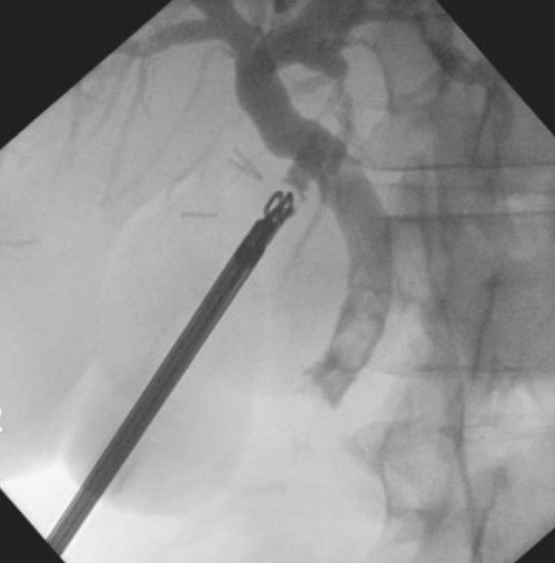
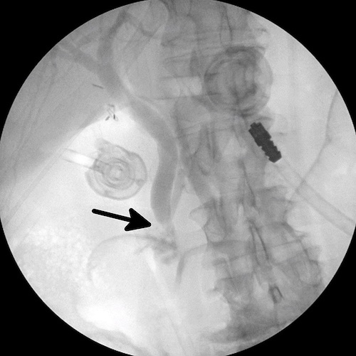
- Findings: Filling Defect or No Drainage into the Duodenum
- Initial Approach: Flush
- Give Glucagon (1.0 mg)
- Wait 2 Minutes
- Flush with 100-200 cc Saline
- Repeat Cholangiogram to Evaluate Clearance
- *Can Repeat A Second Time if Needed
- Options if Fails:
- Common Bile Duct Exploration
- Postoperative ERCP
Common Hepatic Duct Injury
- Findings: Common Bile Duct Fills with No Retrograde Filling of the Common Hepatic Duct
- Similar Image Seen if Cholangiocatheter is Just Advanced Too Far into the Common Bile Duct
- Initial Steps: Position in Trendelenburg, Partially Retract the Cholangiocatheter, and Reimage
- Options if Reimaging Still Fails to See the CHD:
- Convert to an Open Procedure to Better Visualize Anatomy and Evaluate for Injury
- Damage Control – Close and Transfer to a Higher Level of Care
Extraluminal Contrast with No Ductal Filling
- Catheter Possibly Dislodged
- Reevaluate Placement

IOC with Filling Defects – Concerning for Choledocholithiasis 8

IOC with Obstruction at the Ampulla – Concerning for Choledocholithiasis 9
References
- MacFadyen BV. Intraoperative cholangiography: past, present, and future. Surg Endosc. 2006 Apr;20 Suppl 2:S436-40.
- Hope WW, Fanelli R, Walsh DS, Price R, Stefanidis D, Richardson WS. Clinical Spotlight Review: Intraoperative Cholangiography. SAGES. 2017.
- Brunt LM. Should We Utilize Routine Cholangiography? Adv Surg. 2022 Sep;56(1):37-48.
- Temperley HC, O’Sullivan NJ, Grainger R, Bolger JC. Is the use of a routine intraoperative cholangiogram necessary in laparoscopic cholecystectomy? Surgeon. 2023 Jan 27:S1479-666X(23)00003-3.
- Akolekar D, Nixon SJ, Parks RW. Intraoperative cholangiography in modern surgical practice. Dig Surg. 2009;26(2):130-4.
- Velez DR. Cholangiocatheter. The Operative Review of Surgery.
- Al Beteddini OS, Amra NK, Sherkawi E. Ciliated foregut cyst in the triangle of Calot: the first report. Surg Case Rep. 2016 Dec;2(1):20. (License: CC BY 4.0)
- Al-Habbal Y, Reid I, Tiang T, Houli N, Lai B, McQuillan T, Bird D, Yong T. Retrospective comparative analysis of choledochoscopic bile duct exploration versus ERCP for bile duct stones. Sci Rep. 2020 Sep 7;10(1):14736. (License: CC BY 4.0)
- Copeland D, Blears E E, Zhu Z, et al. (February 09, 2019) Novel Technique for Laparoscopic Common Bile Duct Exploration Using Endovascular Instrumentation. Cureus 11(2): e4041. (License: CC BY 3.0)