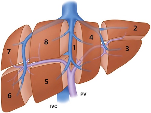
Couinaud Segments 1
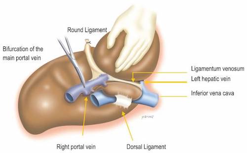
Ligamentum Venosum & Teres 2
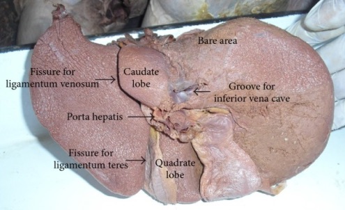
Underside of the Liver Showing Fissures of the Ligamentum Teres & Venosum 3
Portal Triad
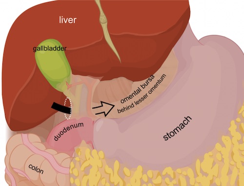
Foramen of Winslow 4
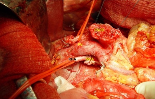
Replaced Right Hepatic Artery (Off SMA) 5
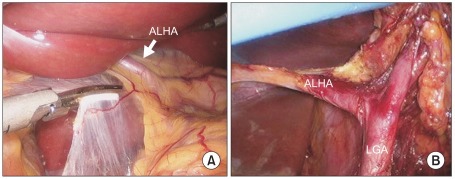
Replaced Left Hepatic Artery (Off Left Gastric) 6
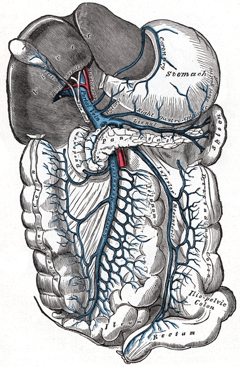
Portal Venous System 7
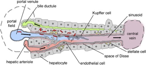
Hepatic Lobule 8
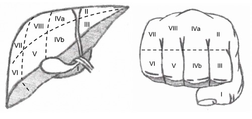
Liver Segments
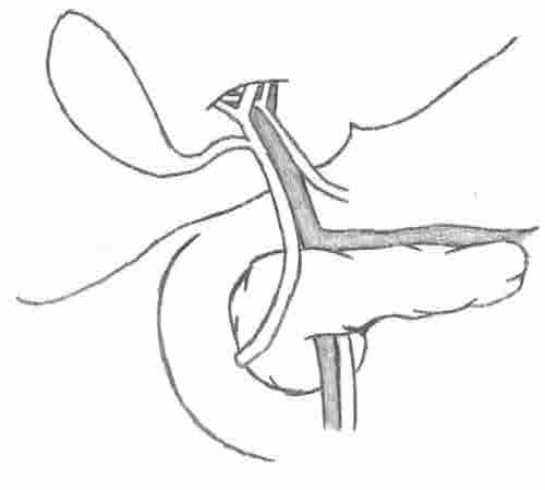
- “Handy” Mnemonic Using Fist & Fingers
Portal Triad Orientation
- *P-P: Portal is Posterior
- Bile Ducts are Lateral (From the Gallbladder which is Lateral)
- Proper Hepatic Artery is Medial (From the Aorta which is Medial)