Vascular: Lymphatic Pathology

Lymphedema

Basics

  • Definition: Swelling Due to Accumulation of Lymphatic Fluid
  • From Lymphatic Obstruction or Incompetence
  • Accumulation of Hyaluronan & Glycoproteins Causes Increased Fibroblasts, Keratinocytes & Macrophages
    • Increased Collagen & Collective Tissue Causes Progressive Subcutaneous Fibrosis

Causes

  • Primary Lymphedema Mn
    • Congenital Lymphedema: Present at Birth
    • Lymphedema Praecox: Presents During Childhood & Adolescence
      • Most Common Primary Cause (94%)
    • Lymphedema Tarda: Presents as an Adult
  • Secondary (Acquired) Lymphedema
    • More Common Than Primary
    • Most Common Cause in U.S.: ALND
    • Most Common Cause in the World: Filariasis (Wuchereria bancrofti Parasite)

Presentation

  • Painless Leg Swelling
  • Woody Edema: From Subcutaneous Fibrosis
  • Swelling Extends into the Feet
    • Stemmer Sign – Unable to Pinch Skin of Second Toe
    • Feet Have Characteristic “Buffalo Hump”
    • “Square Toes”
  • Difficult to Distinguish from Venous Stasis

Grading

  • Latent Phase: Fluid Accumulation Around Lymphatics without Edema
  • Grade I: Pitting Edema Relieved by Elevation, No Fibrosis
  • Grade II: Non-Pitting Edema Not Relieved by Elevation, Fibrosis Present
  • Grade III: Lymphostatic Elephantiasis, Irreversible Edema from Repeated Inflammation & Fibrosis

Complications

  • Recurrent Cellulitis
    • Protein-Rich Fluid Provides a Good Substrate for Bacterial Growth
  • Lymphangiosarcoma
    • Presents as Multicentric Raised Blue-Red Nodules
    • Highly Aggressive with Poor Prognosis
    • Metastasizes to Lung Early
    • Stewart-Treves Syndrome: From Chronic Lymphedema
      • Majority Occur Post Mastectomy or ALND
      • Presentation: Dark Purple Lesions (Bruises) of Arm 5-10 Years Later
      • Treatment: Early Wide Local Excision
        • Often Requires Amputation
  • Malnutrition
    • From Severe Loss of Proteins

Diagnosis

  • Most Often a Clinical Diagnosis
  • Strongly Consider Venous US to Rule Out Venous Pathology
  • If Uncertain: Lymphoscintigraphy (Nuclear Medicine Radiotracer)

Treatment

  • Initial Approach: Conservative Management
    • Compression Stockings
    • Elevation
    • Physiotherapy & Manual Lymphatic Decompression (Massage Technique)
    • Skin Care
    • Weight Loss
  • Microsurgery
    • Options:
      • Lymphovenous Anastomosis
      • Vascularized Lymph Node Transfer
    • Indications:
      • Failure of Conservative Management
      • Recurrent Cellulitis
      • Functional Limitation
      • Deformity
      • Refractory Pain
      • Impaired Quality of Life

Lymphedema; (Left) Left Leg, (Right) Right Arm 1

Lymphangiosarcoma 16 Years After Mastectomy 2

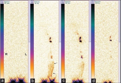
Serial Lymphoscintigraphy Showing Lymphedema ((a) Immediate, (b) 1h, (c) 4h, (d) 24h); RLE No Uptake in Lymph Nodes or Channels, LLE with Dermal Backflow (Arrows) 3

Similar Pathology

  • Venous Stasis
 Venous StasisLymphedema
PittingYesEarly, Not Late
Feet/ToesSparesAffects Mn
HyperpigmentationYesNo
Recurrent CellulitisNoYes
  • Lipedema
    • Symmetric Leg Enlargement from Fat Distribution
    • Similar to Lymphedema but Does Not Affect Feet
    • Tx: Weight Loss

Other Lymphatic Pathology

Lymphangitis

  • Inflammation Along Lymphatic Channels
  • Most Commonly from a Skin Infection
  • Most Common Organisms:
    • Acute Infection: Group A Streptococcus
    • Chronic Infection: Sporothrix schenckii
  • Presentation: Tender Red Linear Streaking
  • Tx: Penicillin
    • Clindamycin if Allergic

Lymphocele (Cystic Lymphangioma)

  • Lymphatic Fluid Collection without an Epithelial Lining
  • Prone to Leakage (No Platelets or Clotting Factors)
  • Most Common Causes: Surgery or Trauma
  • Most Common Site: Groin
  • Treatment: Percutaneous Drainage
    • If Fails: Surgical Resection (Some Prefer as the Primary Treatment)

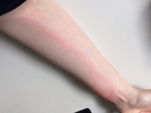
Lymphangitis After Bug Bites 4

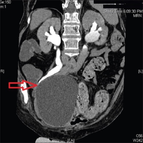
Pelvic Lymphocele After Radical Cystectomy 5

Mnemonics

Presentation of Primary Lymphedema

  • P-P: Praecox in Peds
  • Tardy/Later: Tarda in Adults

Venous Stasis vs Lymphedema

  • Venous – “V” Looks Like a Single Leg WITHOUT the Foot
  • Lymphatic – “L” Looks Like a Single Leg WITH the Foot (Lymphedema Affects the Feet)

References

  1. Cannon S. Pneumatic compression devices for in-home management of lymphedema: two case reports. Cases J. 2009 Mar 23;2:6625.(License: CC BY-3.0)
  2. Sepah YJ, Umer M, Qureshi A, Khan S. Lymphangiosarcoma of the arm presenting with lymphedema in a woman 16 years after mastectomy: a case report. Cases J. 2009 Sep 1;2:6887. (License: CC BY-3.0)
  3. Kalawat TC, Chittoria RK, Reddy PK, Suneetha B, Narayan R, Ravi P. Role of lymphoscintigraphy in diagnosis and management of patients with leg swelling of unclear etiology. Indian J Nucl Med. 2012 Oct;27(4):226-30. (License: CC BY-NC-SA-3.0)
  4. Akinerov. Wikimedia Commons. (License: CC BY-SA-4.0)
  5. Bankar SS, Bakshi GK, Prakash G, Sable NP. Delayed complication of pelvic lymphocele: Ileal conduit obstruction. Indian J Urol. 2015 Jul-Sep;31(3):254-5. (License: CC BY-NC-SA-3.0)