Pancreas: Necrotizing Pancreatitis
Presentation
General
- Pancreatic Tissue Dies Due to Inflammation & Injury
- State of Infection:
- Sterile Initially
- Infected After 2-3 Weeks
- Abscess After 5 Weeks – From Liquefication of Infected Necrosis
- Most Common in Obese
Complications
- Portosplenomesenteric Venous Thrombosis (PSMVT) – 50% Risk
- Diabetic Risk After Necrosectomy: 25%
- Risk Factors: Chronic Pancreatitis & Increased Size Resected
Hemorrhagic Signs Mn
- Cullen Sign: Periumbilical Ecchymosis
- Grey Turner Sign: Flank Ecchymosis
- Fox’s Sign: Ecchymosis Over Inguinal Ligament
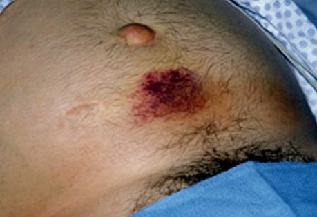
Cullen Sign 1
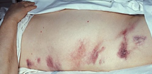
Grey Turner Sign 1
Diagnosis
Diagnosis of Necrosis
- CT: Based on Revised Atlanta Classification
- Revised Atlanta Classification:
- Acute Necrotic Collection
- ≤ 4 Weeks
- Heterogenous Fluid Density – Some Solid Components
- No Defined Wall
- Intrapancreatic or Extrapancreatic
- Walled-Off Pancreatic Necrosis (WOPN)
- > 4 Weeks
- Heterogenous Fluid Density
- Well Defined Wall
- Acute Necrotic Collection
Diagnosis of Infection
- CT Showing Gas within the Necrosis (Highly Specific but Not Sensitive)
- Percutaneous Needle Aspiration Should Be Considered for Prolonged Illness if CT is Not Specific – May Consider Forgoing if Planning to Treat Empirically & Aspiration Will Not Change Management
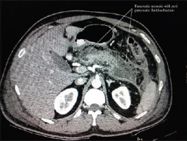
Acute Necrotic Collection 2
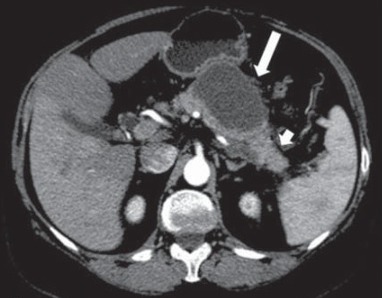
Walled-Off Pancreatic Necrosis 3
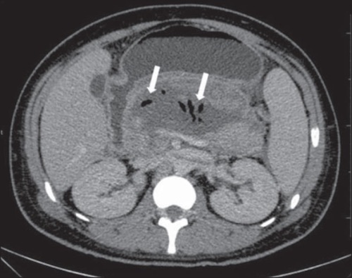
Infected Pancreatic Necrosis 4
Treatment
Sterile Pancreatic Necrosis
- Primary Treatment: Medical Therapy (No Antibiotics Necessary)
- Surgical Debridement:
- Indications:
- Failure to Thrive
- Persistent Abdominal Pain
- Worsening Organ Failure
- Should Be Delayed Until 30 Days After Diagnosis
- Mortality:
- < 15 Days: 75%
- 15-29 Days: 45%
- ≥ 30 Days: 8%
- Indications:
Infected Pancreatic Necrosis
- Stable: “Step-Up Approach”
- Unstable: Antibiotics & Open Necrosectomy
- Use Blunt Finger Dissection – Best to Differentiate Live vs Necrotic Tissue
“Step-Up Approach”
- Approach for Management of Infected Necrotizing Pancreatitis
- Approach:
- Initial: Antibiotics & Drainage
- Antibiotics: Broad Spectrum (Imipenem – Excellent Pancreatic Penetration)
- Drainage: Percutaneous or Endoscopic Approach
- If Fails After 72 Hours: Second Drainage Procedure or “Upsize” the Drain
- If Fails After 72 Hours Again: Video-Assisted Retroperitoneal Debridement (VARD)
- If Still Fails: Laparoscopic/Open Debridement
- Initial: Antibiotics & Drainage
- Delayed Debridement Reduces Mortality & Severe Complications
- Minimally Invasive Debridement Has Lower Risk of Widespread Contamination & Systemic Complications (Diabetes/Multisystem Organ Failure)
- Mortality Unchanged
Mnemonics
Hemorrhagic Signs in Necrotizing Pancreatitis
- Cullen: Think “C” Around the Umbilicus
- Grey-Turner: “Turned” to the Side
- Fox: “Foxy” Lady in the Groin
References
- Wikimedia Commons. (License: CC BY-2.0)
- Pandiaraja J. Another cutaneous sign of acute pancreatitis. Indian J Crit Care Med. 2016 May;20(5):313-4. (License: CC BY-NC-SA-3.0)
- Cunha EF, Rocha Mde S, Pereira FP, Blasbalg R, Baroni RH. Walled-off pancreatic necrosis and other current concepts in the radiological assessment of acute pancreatitis. Radiol Bras. 2014 May-Jun;47(3):165-75. (License: CC BY-NC-3.0)
- Cunha EF, Rocha Mde S, Pereira FP, Blasbalg R, Baroni RH. Walled-off pancreatic necrosis and other current concepts in the radiological assessment of acute pancreatitis. Radiol Bras. 2014 May-Jun;47(3):165-75. (License: CC BY-NC-3.0)