The Majority of Omental Tumors are Metastatic from Other Sites and Represent Advanced Disease
“Milky Spots” of the Omentum Containing Resident Macrophages May Act as Secondary Lymphoid Organs to Mitigate Inflammation and Contribute to the Spread of Malignancy
Tumors Spread by Both Direct and Hematogenous Routes
Primary Omental Tumors – Originate from the Omentum Itself (Rare)
Liposarcoma
Leiomyosarcoma
Hemangiopericytoma
Fibrosarcoma
Mesothelioma
Malignant Fibrous Histiocytoma
Gastrointestinal Stromal Tumor (GIST) – Can Be Primary or Secondary
Desmoid Tumor
Most Common Secondary Omental Tumors – Originate from Another Source and Spread to the Omentum
Uterine
Ovarian
Colorectal
Gastric
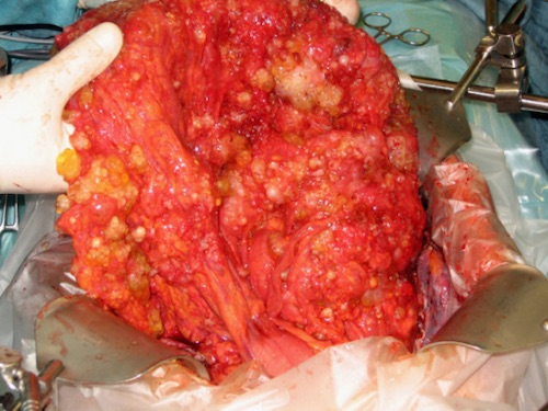
“Omental Caking” Refers to Diffuse Soft Tissue Thickening of the Omentum
Can Be from Metastases or Infection
Often Seen on CT
Most Common Cause: Ovarian Carcinoma
Omental Caking 1
Presentation and Diagnosis
Symptoms
Many Remain Asymptomatic for a Prolonged Period of Time
Abdominal Pain/Discomfort
Increased Abdominal Girth
Palpable Abdominal Mass
Nausea and Vomiting
Ascites
Weight Gain or Weight Loss
CT and MRI Can Evaluate the Tumors and Evaluate for Other Primary Intraabdominal Sources
Definitive Diagnosis is Generally Only Made by Pathologic Examination of the Tumor
Options for Tissue Diagnosis
Fine Needle Aspiration (FNA) – Most Often Inadequate for Tissue Sampling but Accuracy May Be Improved By Following Strict Protocols
Core Needle Biopsy (CNB)
Surgical Resection
Treatment
Management Varies by Tumor Type and Primary Source
Isolated Primary Omental Tumors are Most Often Managed by Surgical Omentectomy (Resection) with or without Chemotherapy
Peritoneal Carcinomatosis: Cytoreductive Surgery (CRS) and HIPEC if Appropriate
Glockzin G, Schlitt HJ, Piso P. Peritoneal carcinomatosis: patients selection, perioperative complications and quality of life related to cytoreductive surgery and hyperthermic intraperitoneal chemotherapy. World J Surg Oncol. 2009 Jan 8;7:5. (License: CC BY-2.0)