Peritoneum: Anatomy and Physiology
Nolan R. King, MD
Table of Contents
Peritoneum
Peritoneum: A Bidirectional Semipermeable Membrane Lining the Abdominal Cavity
Physiology
- Absorbs Blood and Fluid Through Lymphatics
- Made of Simple Squamous Epithelium
Layers
- Visceral Peritoneum: Inner Layer Covering the Abdominal Organs
- Parietal Peritoneum: Outer Layer Covering the Abdominal Wall
Visceral Peritoneum
- Covers the Stomach, Small Intestine, Liver, Spleen, and Transverse Colon
- Few Nerve Fibers – Poor Pain Localization
- The Omentum and Mesentery are Folds of Visceral Peritoneum
- Blood Supply: Splanchnic Vessels
Parietal Peritoneum
- Covers the Abdominal Wall, Pelvis, and Diaphragm
- Blood Supply: Intercostals, Subcostals, Lumbar, and Iliac Vessels
- White Line of Toldt – Lateral Reflections of Parietal Peritoneum Along the Sides of the Ascending and Descending Colon
Peritoneal Cavity: The Potential Space Between the Two Layers – Normally Filled with Minimal Serous Fluid (50 cc)
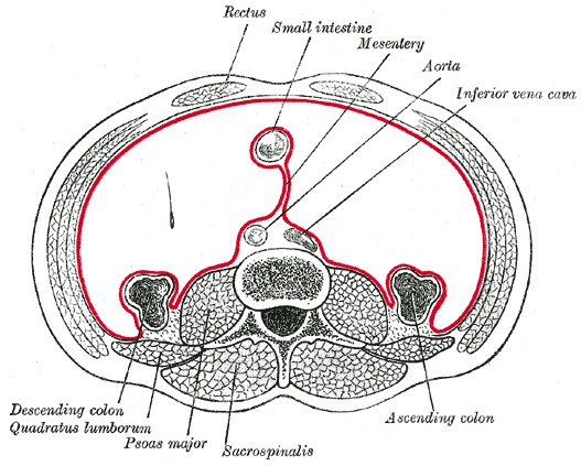
Horizontal Cross-Section of the Abdomen and Peritoneal Covering (Red)
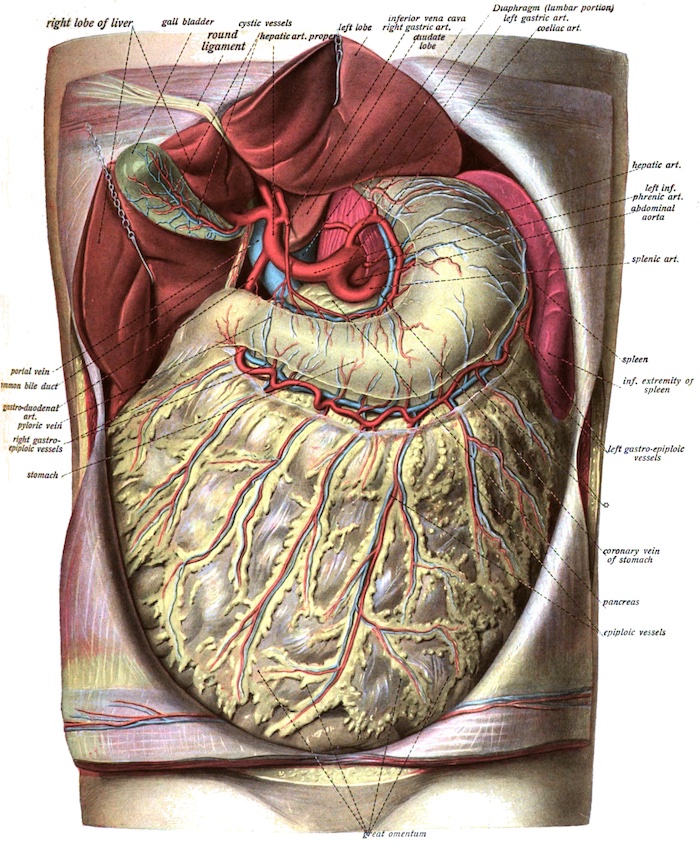
Omentum
Omentum: Large Folds of Visceral Peritoneum Containing Adipose Tissue
Structures
- Lesser Omentum: Hangs off the Lesser Curvature of the Stomach
- Greater Omentum: Hangs off the Greater Curvature of the Stomach
Lesser Omentum
- Also Known as the “Gastrohepatic Omentum”
- Hangs Down from the Liver to the Lesser Curvature of the Stomach and Duodenum
- Contains Multiple Ligaments:
- Hepatogastric Ligament – Liver to Stomach
- Hepatoduodenal Ligament – Liver to Duodenum
- Blood Supply: Right and Left Gastric Arteries
Greater Omentum
- Also Known as the “Gastrocolic Omentum”
- Extends From the Greater Curvature of the Stomach to Cover the Small Intestine Before Doubling Back and Ascending to the Transverse Colon
- Larger than the Lesser Omentum
- Contains Multiple Ligaments:
- Gastrophrenic Ligament – Stomach to Left Diaphragm
- Gastrocolic Ligament – Stomach to Transverse Colon
- Gastrosplenic Ligament – Stomach to Spleen
- Blood Supply: Right and Left Gastroepiploic Arteries
- Considered to Have a Rich Vascular Supply
Functions
- Immune-Regulation:
- Adheres to Sites of Inflammation and Expands to Contain Intraperitoneal Infection
- “Milky Spots” Within the Omentum Contain Resident Macrophages to Allow Rapid Migration to Sites of Inflammation for Bacterial Clearance
- Tissue Regeneration:
- Omentum Promotes Wound Healing with Tissue Regeneration
- Promotes Neovascularization/Neoangiogenesis in Adjacent Structures
- Biological Mechanism is Poorly Understood
- Contains Double the Normal Concentration of Tissue Factor – Promotes Extrinsic Coagulation and Wound Isolation in Trauma/Infection
Omental Transposition/Flap Over an Injured Organ/Tissue Can Be Used to Promote Healing (Graham Patch)
Greater Omentum
Mesentery
Mesentery: A Double Fold of Visceral Peritoneum Extending from the Posterior Abdominal Wall to the Intestine
The Mesentery is a Single Structure (An Internal Organ) Extending from the Duodenojejunal Flexure to the Distal Mesorectum – Not Separate Sections with Separate Insertions
Structures
- Mesentery Proper – Mesentery to the Small Intestine (Jejunum/Ileum)
- “Root of the Mesentery” – The Point Where the Mesentery Proper Attaches to the Posterior Wall
- Transverse Mesocolon – Mesentery to the Transverse Colon
- Sigmoid Mesocolon – Mesentery to the Sigmoid Colon
- Mesoappendix – Mesentery Extending from the Distal Mesentery Proper to the Appendix
- Mesorectum – Mesentery to the Upper Third of the Rectum
Functions
- Anchors Abdominal Organs to the Posterior Abdominal Wall
- Contains the Blood Vessels (SMA, IMA, Portal System) and Nerves that Supply the Intestine
- Numerous Lymphatic Vessels and Lymph Nodes
- Stores Fat
Mesentery Proper 1
Retroperitoneum
Retroperitoneum: The Anatomical Space Behind the Peritoneum
Retroperitoneal Organs Have Peritoneum Only on the Anterior Surface and are Not Suspended by Mesentery
Retroperitoneal Structures
- GI Tract
- Esophagus
- Duodenum – Not the First Part
- Colon (Ascending and Descending) – Not the Transverse or Sigmoid
- Rectum
- Aorta and Inferior Vena Cava (IVC)
- Pancreas – Not the Tail
- Kidneys
- Ureters
- Suprarenal/Adrenal Gland
- MNEMONIC: “SAD PUCKER” (Suprarenal, Aorta/IVC, Duodenum, Pancreas, Ureters, Colon, Kidneys, Esophagus, Rectum)
Origin
- Primary Retroperitoneal: Structures Retroperitoneal Throughout Development
- Esophagus
- Rectum
- Aorta and Inferior Vena Cava (IVC)
- Kidneys
- Ureters
- Suprarenal/Adrenal Gland
- Secondary Retroperitoneal: Structures Were Initially Intraperitoneal and Suspended by Mesentery by Migrated Retroperitoneal During Development
- Duodenum – Not the First Part
- Colon (Ascending and Descending) – Not the Transverse or Sigmoid
- Pancreas – Not the Tail
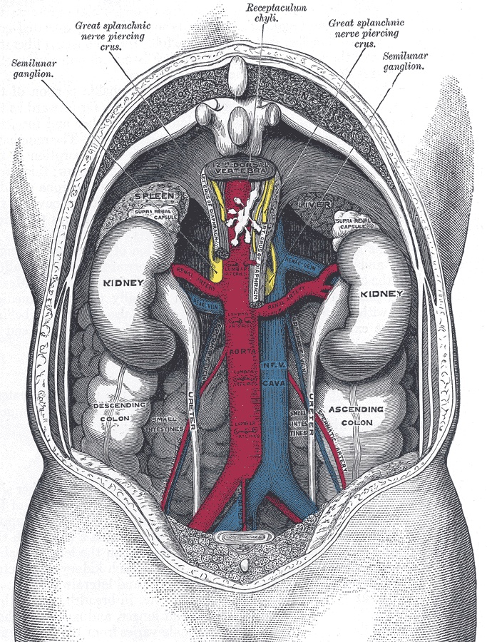
Retroperitoneum – Viewed from Behind
References
- https://www.scientificanimations.com/wiki-images (License: CC BY-SA-4.0)