
Rectus Sheath Hematoma (RSH)
Rectus Sheath Hematoma (RSH)
David Ray Velez, MD
Table of Contents
Definition
Definition: Accumulation of Blood within the Rectus Sheath
Etiology and Epidemiology
The Majority are Due to Injury to a Branch of the Epigastric Vessels
The Inferior Epigastric Vessels are Particularly Susceptible to Injury Because Their Relatively Fixed Course within the Rectus Sheath Limits Mobility – Increasing Vulnerability to Shearing Forces
Most Are Spontaneous and Not from Trauma
Risk Factors
- Anticoagulation and/or Antiplatelet Use – Present in 50-89% of Patients
- Elderly (Smaller Muscle)
- Female Sex
- Pregnancy
- Asthma/COPD – Vigorous Coughing Spells
- Chronic Medical Conditions (HTN, Cirrhosis, CKD)
- Abdominal Injections – Particularly Subcutaneous Heparin/Lovenox
Presentation and Diagnosis
Presentation
- Abdominal Pain
- Abdominal Mass/Bruising
- Nausea and Vomiting
- Fothergill’s Sign – Does Not Cross Midline and Does Not Change with Flexion
- Hernias are Generally More Painful/Prominent with Rectus Flexion
- Carnett’s Sign – Point of Maximal Tenderness Does Not Change from Supine to Sitting
Complications
- Hemorrhagic Shock
- Infection
- Abdominal Compartment Syndrome
Diagnosis
- Generally Diagnosed by CT Imaging
- May Also Consider US or MRI in Select Scenarios
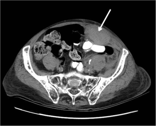
Rectus Sheath Hematoma on CT 1
Classification
Type I: Small, Confined to the Rectus, and Does Not Cross Midline
Type II: Confined to the Rectus but Crosses Midline
- Dissects Along Transversalis Fascial Plane
Type III: Large and Not Confined
- Usually Below the Arcuate Line – May Cause Severe Bleeding Due to Lack of Aponeurosis to Contain/Tamponade
- Often See Hemoperitoneum and/or Blood in Prevesical Space of Retzius
Management
The Majority are Managed Conservatively with Observation and Medical Therapy
Reverse Anticoagulation if Unstable or with Active Bleeding
Indications for IR Angioembolization:
- Hemodynamically Unstable
- Ongoing Requirement for Blood Transfusions
- Enlarging/Expanding
- Active Contrast Extravasation on CT
Indications for Surgery:
- Skin Necrosis
- Failure of Angioembolization
- Compressive Symptoms (Femoral Neuropathy, etc.)
- Abdominal Compartment Syndrome – Rare
Surgical Intervention
- Procedure: Surgical Evacuation and Vessel Ligation
- Incision: Midline or Paramedian to Expose the Posterior Sheath
- Avoid Entering the Peritoneum
- Obtain Hemostasis by Electrocautery and Ligation of Any Bleeding Vessels
- May Require Ligation of the Superior or Inferior Epigastric Vessels – Generally Well Tolerated Due to Good Collateral Flow
- Pack with Wet-to-Dry Dressings for Compression or Close Space Over a Drain
Inferior Epigastric Artery Ligation
- Incision: Oblique Over the Groin
- Carried Down to the Inguinal Ligament
- Inferior Epigastric Found Branching Off the Medial Aspect of the Distal External Oblique Artery
- Rarely Required Only for Persistent Bleeding that Cannot Be Controlled
References
- Sullivan LE, Wortham DC, Litton KM. Rectus sheath hematoma with low molecular weight heparin administration: a case series. BMC Res Notes. 2014 Sep 1;7:586. (License: CC BY-2.0)