Vascular: Carotid Artery Stenosis

Carotid Artery Stenosis

Basics

  • Most Common Site: Bifurcation

Presentation

  • Amaurosis Fugax – Transient Vision Changes to Ipsilateral Eye
    • From Occlusion of the Ophthalmic Artery (First Branch of ICA)
    • Cholesterol Embolus (Hollenhorst Plaque) Seen on Eye Exam
  • Transient Ischemic Attacks (TIA)
  • Ischemic Stroke

Characteristics with Increased Risk of Stroke

  • Plaque Characteristics:
    • Diameter/Area
    • Progression
    • Echolucent
    • Ulceration
    • Fibrous Cap Disruption
    • Inflammation by MRI
  • Patient Characteristics:
    • Neurologic Symptoms
    • Contralateral Carotid Occlusion (CCO)
    • Past Contralateral Stroke
    • Renal Insufficiency
    • Smoking
    • Clinically Silent Emboli

General Risk of Stroke (ACAS/NASCET Study Findings)

  • Asymptomatic > 60% Stenosis:
    • Medical Therapy: 11% at 5 Years
    • CEA: 5.1% at 5 Years
  • Symptomatic 50-69% Stenosis:
    • Medical Therapy: 22.2% at 2 Years
    • CEA: 15.7% at 2 Years
  • Symptomatic > 70% Stenosis:
    • Medical Therapy: 26% at 2 Years
    • CEA: 9% at 2 Years

Carotid Plaque 1

Hollenhorst Plaque 2

Diagnosis

  • First Line Imaging Modality: Duplex US
    • Good for Stenosis 70-99% But Less Sensitive for Stenosis 50-69%
    • Based Primarily on Velocities Opposed to Visualization of the Physical Plaque
  • Ultrasound Criteria (Society of Radiologists in Ultrasound):
Degree of Stenosis Primary Parameters Secondary Parameters
ICA PSV Plaque Estimate ICA/CCA PSV Ratio ICA EDV
Normal < 125 cm/s None < 2.0 < 40 cm/s
< 50% < 125 cm/s < 50% < 2.0 < 40 cm/s
50-69% 125-230 cm/s ≥ 50% 2.0-4.0 40-100 cm/s
> 70% > 230 cm/s ≥ 50% > 4.0 > 100 cm/s
Near Occlusion High, Low or Undetectable Visible Variable Variable
Total Occlusion Undetectable Visible with No Detectable Lumen Not Applicable Not Applicable
    • *ICA – Internal Carotid Artery, CCA – Common Carotid Artery, PSV – Peak Systolic Velocity, EDV – End Diastolic Velocity
  • CTA Indicated if Concern for Anatomy:
    • High Bifurcation
    • Reoperative CEA
    • Planned Carotid Stenting – US Alone Not Sufficient
  • Gold Standard: Angiography

Normal ICA on Duplex US 3

ICA Stenosis on Duplex US 3

ICA Stenosis on Angiography 3

Treatment

General Treatment Approach

  • Asymptomatic:
    • < 80% Stenosis: Medical Therapy
    • 80-99% Stenosis: Carotid Endarterectomy (CEA) vs Carotid Artery Stenting (CAS)
      • *Cutoff is Debated and Older Major Studies Had Recommended 60%
  • Symptomatic:
    • < 50% Stenosis: Medical Therapy
    • 50-99% Stenosis: Carotid Endarterectomy (CEA) vs Carotid Artery Stenting (CAS)
    • Timing Post CVA:
      • Fluctuating Neuro Status & Evolving TIA: Emergent
      • TIA or Non-Disabling: < 2 Weeks
        • Too Early (< 24 Hours) – Risk Reperfusion Injury
      • Moderate-Large Stroke or Midline Shift: 4-6 Weeks
        • Higher Reperfusion Risk
      • Hemorrhagic Stroke: 6-8 Weeks
      • Completely Disabling: None
  • 100% Occlusion: Anticoagulation
    • Do Not Open, Risk Reperfusion Injury
  • May Consider Repair at a Lower Percentage if Ulcerated (Higher Risk)

Medical Therapy

  • Antihypertensive Therapy
  • Statin Therapy
  • Antiplatelet Therapy
  • Smoking Cessation

Carotid Endarterectomy (CEA)

  • Stenosis Benefit of Major Studies:
    • Symptomatic:
      • NASCET: > 70%; > 50% to Lesser Degree
      • ECST: > 60%
    • Asymptomatic:
      • ACAS: > 60%
      • ACST: > 60%
      • *Medical Managements were Outdated and Cutoff more Debated – Some Recommend as High as 80%
  • Contraindications:
    • Poor Baseline Functional Status with No Benefit
    • Short Life Expectancy
  • Less Benefit in Women & Asymptomatic Disease

Carotid Artery Stenting (CAS)

  • Potential Indications:
    • “Hostile Neck” (Effected by Prior Radiation, Stoma or Surgery)
    • Recurrent Stenosis
    • High Lesions (Above C2 Vertebrae) – Avoid Mandibular Subluxation Required for Open CEA
    • Low Lesions (Below Clavicle)
  • Potential Contraindications:
    • Symptomatic Disease – Inferior Outcomes to CEA
    • Vessel Tortuosity
    • Adverse Lesion Characteristics:
      • Length > 15 mm
      • Echolucent
      • Circumferential Lesion
      • Preocclusive Lesion
      • Tandem Lesions
  • Comparison to CEA (CREST Trial):
    • Similar Overall Rates of Stroke, MI & Death
    • CAS Has Higher Rates of Perioperative Stroke (4.1% vs 2.3%)
    • CEA Has Higher Rates of Perioperative MI (2.3% vs 1.1%)

References

  1. Uthman E. Wikimedia Commons. (License: CC BY-2.0)
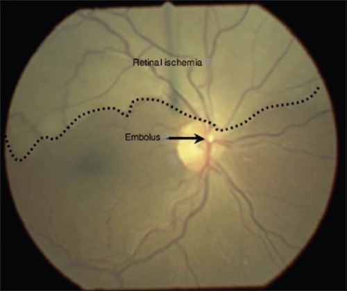
  2. Jones RG, Peall A. Sudden unilateral visual field loss. J Emerg Trauma Shock. 2009 Sep;2(3):211-2. (License: CC BY-NC-SA-3.0)
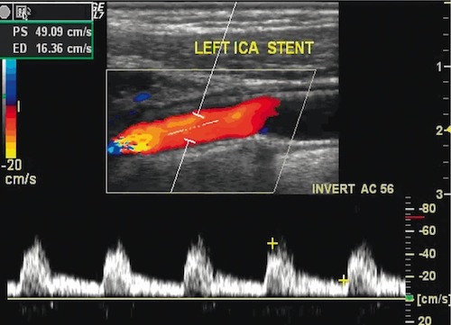
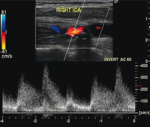
  3. Szczerbo-Trojanowska M, Jargiełło T, Drelich-Zbroja A. Management of carotid stenosis. History and today. J Ultrason. 2013 Mar;13(52):6-20. (License: CC BY-NC-ND-3.0)