General Abdomen: Peritoneal Carcinomatosis
Peritoneal Carcinomatosis
General
- Peritoneum Covered in Carcinomatous Masses
- CT Shows Organ Scalloping
Pseudomyxoma Peritonei
- Mucus & Cystic Masses
- Subset of Peritoneal Carcinomatosis
- Source: Appendix, Small Intestine & Ovary
- Most Common Source: Mucinous Cystadenoma of Appendix
- Most Common Primary Peritoneal Malignancy: Mesothelioma
- More Common in Women
Symptoms
- Increasing Abdominal Girth #1
- Inguinal Hernia #2
- Weight Loss
Peritoneal Cancer Index (PCI)
- Grading Index Used for Research and to Objectively Describe Extent
- Add Largest Lesion Size (0-3) in Each of 13 Regions
- Scores Range 0-39
- Regions:
- Region 0: Central
- Region 1: RUQ
- Region 2: Epigastrium
- Region 3: LUQ
- Region 4: Left Flank
- Region 5: LLQ
- Region 6: Pelvis
- Region 7: RLQ
- Region 8: Right Flank
- Region 9: Upper Jejunum
- Region 10: Lower Jejunum
- Region 11: Upper Ileum
- Region 12: Lower Ileum
- Lesion Size Score:
- 0 = No Tumor
- 1 = ≤ 0.5 cm
- 2 = ≤ 5.0 cm
- 3 = > 5.0 cm
Completeness of Cytoreduction Score
- Scoring System Used to Grade How Complete Cytoreduction Was
- Scores:
- CC-0: No Disease
- CC-1: ≤ 0.25 cm
- CC-2: ≤ 2.5 cm
- CC-3: > 2.5 cm
Prevention of Port Site Metastases
- Reduced Tissue Trauma & Instrument Exchanges
- Minimize Tumor Manipulation
- Use of Protective Retrieval Bags
- Use of Povidone-Iodine to Irrigate Trocar Sites & Rinse Trocar Tips
- Remove Intraabdominal Fluid Before Trocar Removal
- Desufflate Pneumoperitoneum Before Trocar Removal
- Avoid CO2 Leaks & Sudden Desufflation
Treatment
- Extraperitoneal Mets: Systemic Chemotherapy
- No Extraperitoneal Mets: Cytoreductive Surgery & HIPEC
- Abdominal Wall Masses Should Be Excised with the Associated Peritoneum
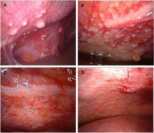
Peritoneal Carcinomatosis on Laparoscopy 1
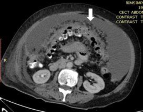
CT Showing Organ Scalloping (Arrow) of Peritoneal Carcinomatosis 2
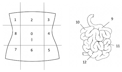
Peritoneal Cancer Index (PCI) Diagram
Intraperitoneal Chemotherapy
HIPEC (Hyperthermic Intraperitoneal Chemotherapy)
- Heated Chemotherapeutic Drugs Infused into Peritoneal Cavity
- Avoids Systemic Circulation Minimizing Toxicity
- Heating Increases Penetration & Cytotoxicity
- Most Common Agents: Oxaliplatin, Cisplatin, Doxorubicin & Mitomycin-C
- Delivery
- Preformed After Surgical Debulking
- Same Operation (Adhesions Create Barrier Preventing Uniform Distribution)
- Before Any Reconstruction or Diversion (Allow Exposure of All Resection Lines)
- Duration: 30-120 Minutes (90 Minutes Common)
- Abdomen is Temporarily Closed
- “Shake & Bake” During Infusion
- Requires No Residual Tumors > 2 mm (Chemo Unable to Penetrate) Mn
- Goal CC-0 or CC-1
- Contraindications:
- Absolute:
- Not Amenable to Complete Cytoreduction
- Poor Performance Status
- Disease Progression on Systemic Therapy
- Malignant Small Bowel Obstruction
- Relative:
- Short-Disease Free Interval – If Metachronous
- Peritoneal Cancer Index > 20
- Serous Ascites
- Absolute:
- Chemotherapy Given Via a Catheter or Subcutaneous Port
- Not Heated
- Administered on Postoperative Day #1-5 After Surgical Debulking
- Able to Give Multiple Cycles with Longer Dwell Time
- Greater Risk of Systemic Absorption – HIPEC Typically Favored
Mnemonics
Maximum Residual Tumor Size in HIPEC
- Nothing Over 2 mm to Move onto 2nd Step
References
- Touboul C, Vidal F, Pasquier J, Lis R, Rafii A. Role of mesenchymal cells in the natural history of ovarian cancer: a review. J Transl Med. 2014 Oct 11;12:271. (License: CC BY-4.0)
- Singh S, Devi YS, Bhalothia S, Gunasekaran V. Peritoneal Carcinomatosis: Pictorial Review of Computed Tomography Findings. International Journal of Advanced Research. 2016;4(7):735–748. (License: CC BY-4.0)