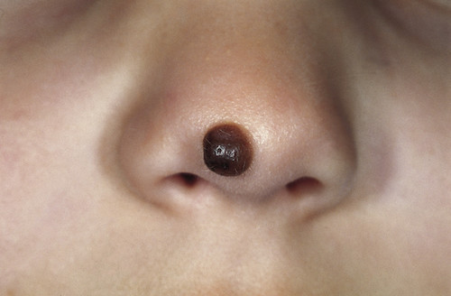
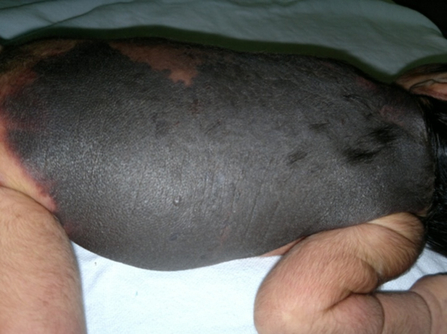
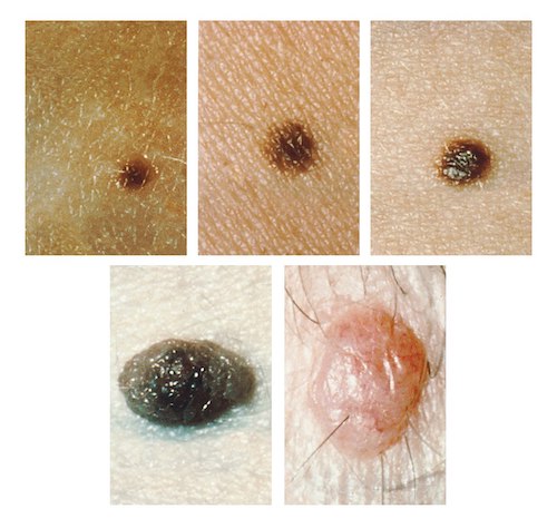
Skin & Soft Tissue: Melanocytic Nevus (Mole) Melanocytic Nevus (Mole) DefinitionsMelanocytic Nevus: Benign Proliferation of Nevus Cells (Type of Melanocyte Clustered in Nests)Congenital Melanocytic Nevus: Present at Birth or First 6 Months of AgeExtend Deeper than Acquired Melanocytic NeviAcquired Melanocytic Nevus: Appear After 2 Years of Age Congenital Melanocytic Nevus (CMN) PresentationPigmented LesionMacules or Slightly Raised PapulesSharply Demarcated BordersGrow Proportionally with the Child – Cover the Same Relative Surface Area Throughout GrowthOccur in Any LocationGiant Lesions May Have a “Garment”/“Bathing Trunk”/“Coat Sleeve” Appearance Due to Large Surface Area Resembling ClothingLarge-Giant Lesions Often Have Other Surrounding Satellite LesionsClassificationSize:Small: < 1.5 cmMedium: > 1.5 cmM1: 1.5-10 cmM2: 10-20 cmLarge: > 20 cmL1: 20-30 cmL2: 30-40 cmGiant: > 40 cmG1: 40-60 cmG2: > 60 cm*Based on the Projected Largest Diameter Achieved by AdulthoodEstimated Size Increase:Head: 1.7xTrunk/Arms: 2.8xLegs: 3.3xSatellite Lesions (Used to Further Classify Large-Giant Lesions):S1: 0S2: 1-19S3: 20-50S4: > 50Melanoma RiskSmall-Medium Sized: < 1%Large-Giant: 2-5%Half of the Risk is During the First Five Years of LifeDiagnosisMostly Clinical (Based on History and Physical Exam)May Consider Skin Biopsy to Rule Out MalignancyTreatmentSmall-Medium: Periodic MonitoringExcision Indications:Difficult Site to Monitor for ChangesParent Preference (Cosmesis or Anxiety)Large-Giant: Surgical ExcisionMay Be Difficult-Impossible to Completely ExciseMay Require Skin Grafting Congenital Melanocytic Nevus 1 Giant Congenital Melanocytic Nevus 2 Acquired Melanocytic Nevus (AMN) Presentation/TypesCommon (Banal)Pigmented LesionSymmetricHomogenous SurfaceRound with Sharply Demarcated BordersSmall (≤ 6 mm)AtypicalShare Clinical Features of Melanoma (Asymmetric, Border Irregular, Color Variation or Diameter > 6 mm)DiagnosisMostly Clinical (Based on History and Physical Exam)May Consider Skin Biopsy to Rule Out MalignancyA Nevus with Different Features from Other Nevi (“Ugly Duckling”) Raises Concern for MelanomaTreatmentCommon (Banal): Periodic Monitoring & Sun ProtectionConsider Excision for Patient Preference (Cosmesis)No Benefit to “Prophylactic” Removal – Most Melanomas Arise De NovoAtypical: Excisional BiopsyRe-Excision of Positive Margins is Debated Acquired Melanocytic Nevus 3 References Sand M, et al. Wikimedia Commons. (License: CC BY-2.0)Sharma S, et al. Wikimedia Commons. (License: GNU FDL)National Cancer Institute. Wikimedia Commons. (License: Public Domain)