
Totally Extraperitoneal (TEP) Repair
Totally Extraperitoneal (TEP) Repair
David Ray Velez, MD
Definition
Definition: A Minimally Invasive Posterior Mesh Repair Performed Entirely Within the Preperitoneal Space without Entering the Peritoneal Cavity
Avoids Intraperitoneal Access While Providing Wide Mesh Coverage of the Entire Myopectineal Orifice
Repairs Both Inguinal Hernias and Femoral Hernias
See Also:
*See Inguinal Hernia
*See Femoral Hernia
*See Inguinal Hernia Repair (IHR)
*See Transabdominal Preperitoneal (TAPP) Repair
Inguinal Anatomy/Laparoscopic Triangles: *See Abdominal Wall: Anatomy
Technique
Infraumbilical Incision
- Make a 1-2 cm Infraumbilical Incision
- Dissect Down to the Rectus Sheath
- Incise the Anterior Rectus Sheath Off Midline
- Bluntly Retract or Split the Rectus Muscle to Access the Posterior Rectus Sheath
Preperitoneal Exposure and Dissection
- Develop the Preperitoneal Space Posterior to the Rectus Muscle
- Can Use Finger Dissection or a Balloon Dissector (Inflates to Spread and Open the Space)
- Insert a 10-12 mm Camera Port Through the Incision into the Preperitoneal Space (Larger to Later Insert the Mesh Through)
- Insufflate the Preperitoneal Space
- Place Two Additional 5-mm Ports Under Direct Visualization Inferiorly Along the Midline
- First ~ 5 cm Below the Camera Port
- Second ~ 5 cm Below the Other (Usually Just Above the Pubis)
- Develop the Space of Retzius Medially – Between the Pubic Symphysis and Bladder
- Develop the Space of Bogros Laterally – Lateral from the Inferior Epigastric Vessels to the ASIS
- Obtain the Critical View of the Myopectineal Orifice (MPO)
Caution: Avoid Inadvertent Violation of the Peritoneal Cavity Which Can Cause Pneumoperitoneum and Loss of the Preperitoneal Working Space – May Consider Veress Needle to Decompress or a Conversion to a TAPP Repair
Repair
- Completely Reduce the Hernia Sac
- Parietalize the Spermatic Cord Structures – Completely Separate the Peritoneum from the Vas Deferens and Gonadal Vessels
- Place a Large Mesh Over the Myopectineal Orifice (MPO)
- Ensure Adequate Circumferential Overlap
- Mesh May Be Left Unfixed or Secured with Tacks, Glue, or Self-Fixating Mesh Depending on Surgeon Preference
- Close Any Inadvertent Peritoneal Tears
Caution: Identify and Avoid Tacks or Sutures within the “Triangles of Danger” (Inferior to the Inguinal Ligament and Lateral to the Vas Deferens)
Closure
- Desufflate Under Direct Visualization While Maintaining Mesh Position
- Remove Ports
- Close the Fascial Defect at the 10-12 mm Port Site
- Close Skin
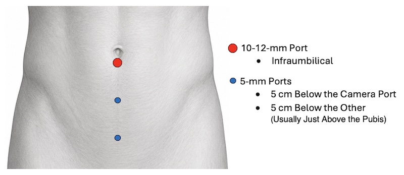
Port Placement
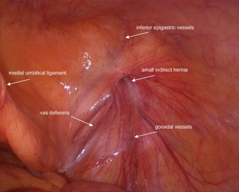
Laparoscopic View of the Inguinal Triangles 1
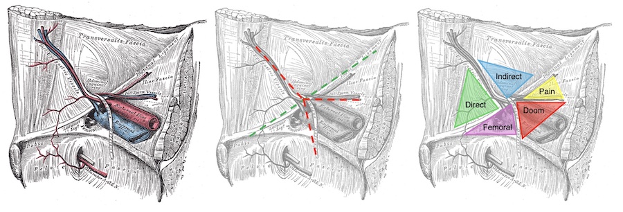
Laparoscopic Inguinal Triangles
Critical View of the Myopectineal Orifice (MPO)
Definition: Complete Exposure of the Myopectineal Orifice (Anatomical Area Viewed) Prior to Mesh Placement During Minimally Invasive Hernia Repair
Similar Concept to the Critical View of Safety in Laparoscopic Cholecystectomy – Although Not Universally Adopted in the Same Way
General Requirements:
- Expose the Pubic Tubercle and Cooper’s Ligament Across the Midline
- Develop the Spaces of Retzius (Medially) and Bogros (Laterally)
- Reduce All Hernia Sacs – Direct, Indirect, Femoral, and Obturator
- Completely Parietalize the Cord Structures
- Reduce Any Cord Lipoma
- Visualize the Psoas Muscle and Iliac Vessels Laterally
- Achieve Complete Hemostasis
- Place a Large Flat Mesh to Cover the Entire Myopectineal Orifice Without Folds or Creases
9 Steps to Establish
- Step 1: Expose the Pubic Tubercle and Cooper’s Ligament Across the Midline
- Step 2: Rule Out a Direct Hernia
- Visualize the Entire Hasselbach Triangle
- Step 3: Develop the Space of Retzius Medially
- Dissect at Least 2 cm Between Cooper’s Ligament and the Bladder
- Facilitates Flat Placement of the Medial Mesh to Avoid Mesh Displacement Caused by Bladder Distention
- Step 4: Rule Out a Femoral Hernia
- Visualize the Femoral Canal Between Cooper’s Ligament and the Iliac Vein
- Step 5: Parietalize the Cord’s Elements
- Completely Separate the Peritoneum from the Cord Structures Until the Cord’s Elements Lie Flat and No Movement of the Cord is Triggered by Pulling the Sac and Peritoneum Upward
- Step 6: Reduce Any Cord Lipomas
- Step 7: Develop the Space of Bogros Laterally
- Extend Beyond the Anterior Superior Iliac Spine (ASIS)
- Step 8: Ensure the Mesh and Any Fixation Lie Above the Inter-ASIS Line
- Avoids Recurrence and Nerve Injury
- Step 9: Place a Large Flat Mesh Only After Complete Dissection and Hemostasis
- Typically ≥ 15 x 10 cm
- Ensure No Creases or Folds
References
- Carter J, Duh QY. Laparoscopic repair of inguinal hernias. World J Surg. 2011 Jul;35(7):1519-25. (License: CC BY-ND-4.0)