
Achalasia
Achalasia
Yaiel G. Rodríguez-Avilés, MD
Table of Contents
Definition
Definition: Primary Esophageal Motility Disorder Characterized by Impaired Relaxation of the Esophagogastric Junction (EGJ) and Absent Peristalsis
Epidemiology
- Incidence: Approximately 1.6 per 100,000 Annually
- Most Commonly Diagnosed Between Ages 25-60
- Affects Males and Females Equally
Pathogenesis and Causes
Pathogenesis
- Selective Loss or Dysfunction of Inhibitory Myenteric (Auerbach) Plexus Neurons in the Distal Esophagus and Lower Esophageal Sphincter (LES)
- Produces an Imbalance Between Excitatory and Inhibitory Input
- Results in Failure of LES Relaxation and Loss of Esophageal Peristalsis
- In Later Stages of the Disease, the Distal Obstruction and Retention of Food and Secretions Can Lead to Esophageal Dilation and Compensatory Remodeling of the Esophageal Wall
Causes
- Primary Achalasia
- Most Cases are Idiopathic
- Proposed Mechanisms:
- Genetic Susceptibility (e.g., Alstrom Syndrome, Down Syndrome)
- Viral Trigger (e.g., HSV-1, HPV, Measles)
- Immune-Mediated Destruction of Inhibitory Neurons
- Secondary Achalasia/Achalasia-Like Disorders
- Pseudoachalasia
- Most Commonly Caused by Malignancy at the EGJ or Gastric Cardia
- May Result from Direct Neural Invasion or Mechanical Obstruction
- Suspected in Patients > 60 Years Old with New-Onset, Rapidly Progressive Dysphagia, and Weight-Loss of < 6 Months Evolution
- Chagas Disease
- Caused by Trypanosoma cruzi
- Produces Destruction of Enteric Ganglion Cells
- May Also Cause:
- Megacolon
- Cardiac Disease
- Other associated disorders
- Amyloidosis
- Sarcoidosis
- Neurofibromatosis
- Multiple Endocrine Neoplasia Type 2B
- Pseudoachalasia
- Opioid-Associated Esophageal Dysfunction
- Can Produce Type III Achalasia or Other Obstructive/Spastic Manometric Patterns
- When Feasible, Perform Manometry After Withholding Opioids for an Appropriate Period
Clinical Presentation
Clinical Presentation
- Dysphagia
- Progressive Dysphagia to Both Liquids and Solids
- Regurgitation
- Regurgitation of Undigested Food and Saliva
- Worse When Supine
- Often Misinterpreted as Vomiting Which May Lead to Evaluation of Gastric Pathologies Rather than an Esophageal Disorder
- Weight Loss
- Usually Mild and Progressive in Primary Achalasia
- Rapid and Severe Weight Loss Should Raise Concern for Pseudoachalasia
- Substernal Chest Pain
- Heartburn (Retrosternal Burning)
- May Be Mistaken for Gastroesophageal Reflux Disease (GERD)
- True GERD Can Also Develop, Particularly After Treatment
- Difficulty Belching (Abelchia)
- Paradoxical Increase in Upper Esophageal Sphincter (UES) Pressure Without a Belch
- Associated Chest Pain and Distention May be Worsened by This “Trapped” Air
Additional Findings
- Nocturnal Cough
- Aspiration and Recurrent Pneumonia
- Hiccups
- Globus Sensation
Diagnosis
Diagnosis
- High-Resolution Manometry (HRM) is the Diagnostic Gold Standard Test
- Esophagogastroduodenoscopy (EGD) Should Be Performed in all Patients with Suspected Achalasia to Exclude Mechanical Obstruction, Malignancy, or Pseudoachalasia
- Timed Barium Esophagram (TBE) is Complementary but Not Diagnostic Itself
- Functional Lumen Imaging Probe (FLIP) if HRM is Inconclusive, Unable to Be Performed, or as Supportive Testing When the Diagnosis Remains Uncertain
High-Resolution Manometry (HRM)
- The Gold Standard Diagnostic Test
- Findings:
- Elevated Median Integrated Relaxation Pressure (IRP)
- Absence of Normal Peristalsis
Timed Barium Esophagram (TBE)
- Classic Findings:
- Dilated Esophagus
- Impaired/Delayed Barium Emptying
- Smooth Tapering at the EGJ
- “Bird’s-Beak” Appearance
- Other Findings:
- Air-Fluid Level
- Absence of a Gastric Air Bubble
- Megaesophagus
- Tortuous or Sigmoid-Shaped Esophagus
- Epiphrenic (Pulsion) Diverticulum
- Can Also Be Used to Objectively Assess Esophageal Emptying and Response to Treatment
Upper Endoscopy/Esophagogastroduodenoscopy (EGD)
- Should Be Performed in All Patients with Suspected Achalasia
- May Be Normal in Achalasia and is Not Diagnostic
- Primary Purpose:
- Exclude Malignancy
- Exclude Stricture or Another Mechanical Obstruction
- Evaluate for Pseudoachalasia
- Findings that Support Achalasia:
- Dilated Esophagus
- Retained Saliva, Liquid, Pills, or Food
- Puckered or Rosette-Like EGJ
- Contracted LES that Can Usually Be Traversed with Gentle Pressure
- Mucosal Biopsy Does Not Establish Achalasia Given that the Primary Abnormality is Within the Myenteric Plexus
Functional Lumen Imaging Probe (FLIP)
- Generally Used When Achalasia is Suspected but HRM is Inconclusive or Unable to Be Performed
- Uses Impedance Planimetry by Balloon Catheter to Measure Cross-Sectional Area and Intra-Luminal Pressure
- The Ratio of the Two Measurements at the Level of the EGJ is Called the Distensibility Index (EGJ-DI)
- Reduced EGJ-DI Supports EGJ Outflow Obstruction and is Consistent with Achalasia in the Appropriate Clinical Context
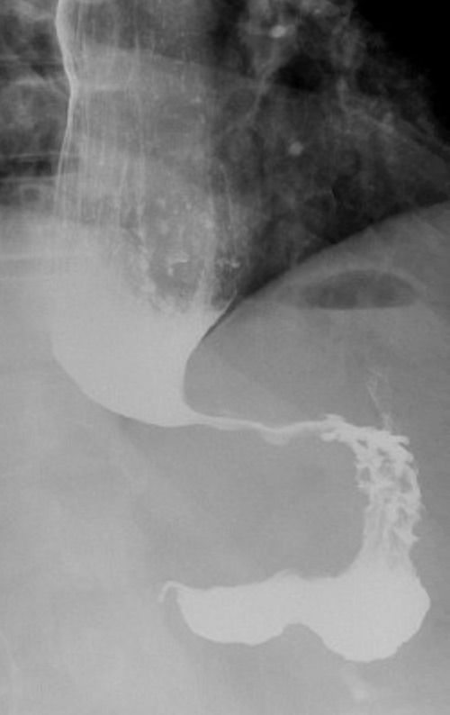
“Bird’s Beak” on Esophagram
Classification/Types
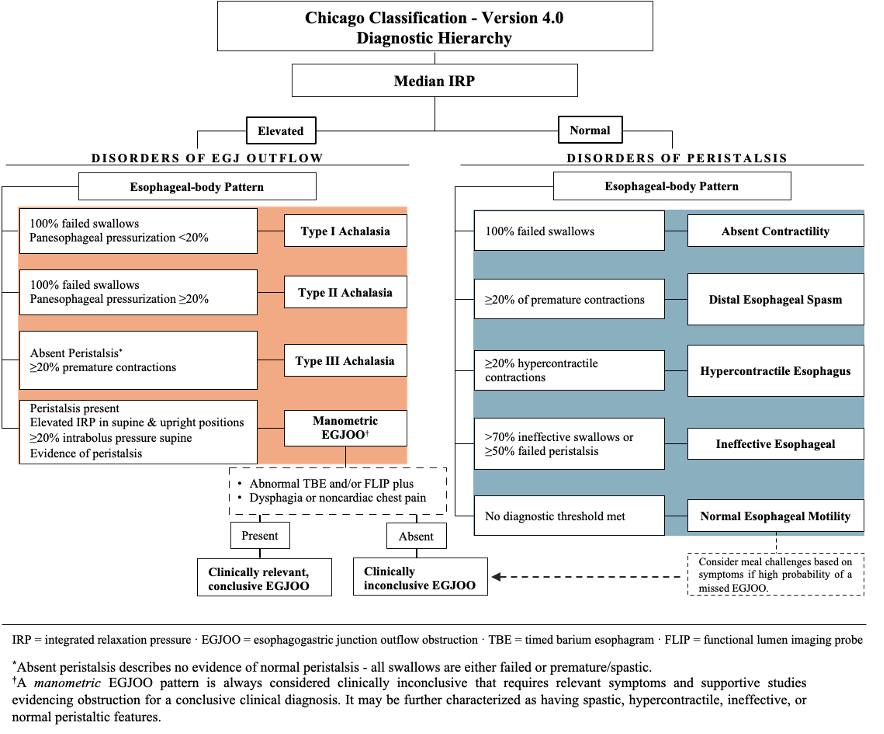
Type I Achalasia (Classic Achalasia)
- Elevated Median Integrated Relaxation Pressure (IRP)
- 100% Failed Peristalsis
- Panesophageal Pressurization (PEP) in < 20% of Swallows
Type II Achalasia
- Elevated Median Integrated Relaxation Pressure (IRP)
- 100% Failed Peristalsis
- Panesophageal Pressurization (PEP) in ≥ 20% of Swallows
Type III Achalasia (Spastic Achalasia)
- Elevated Median Integrated Relaxation Pressure (IRP)
- Absent Peristalsis
- All Swallows are Either Failed or Premature/Spastic
- Premature/Spastic Contractions in ≥ 20% of Swallows
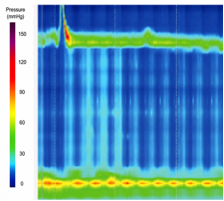
Achalasia Type I: No Contractility with Incomplete LES Relaxation
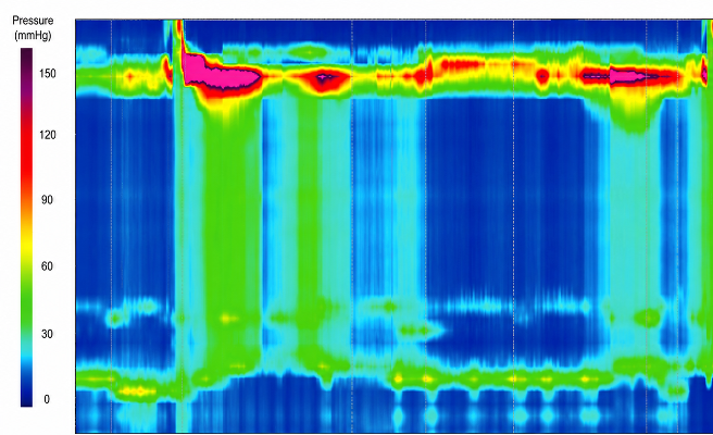
Achalasia Type II: Penesophageal Pressurization (PEP) Bands – Vertical Green Columns
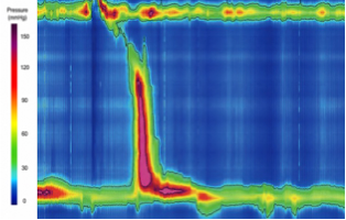
Achalasia Type III: Premature Spastic Contractions
Treatment
General Approach
- Definitive Management:
- Heller Myotomy (HM)
- Peroral Endoscopic Myotomy (POEM)
- Pneumatic Dilation (PD)
- Options if Unable to Tolerate Definitive Therapy:
- Endoscopic Botox Injection into the Lower Esophageal Sphincter (LES)
- Pharmacologic Treatments
Heller Myotomy (HM)
- Procedure: Complete Division of Both the Circular and Longitudinal Muscle Fibers of the Distal Esophagus and Proximal Stomach to Fully Disrupt the Non-Relaxing Lower Esophageal Sphincter (LES)
- Preserves the Inner Submucosa and Mucosa
- Myotomy Extent:
- Proximal: 4-6 cm onto the Distal Esophagus
- Distal: 2-3 cm onto the Gastric Cardia
- Usually Combined with a Partial Fundoplication (Dor or Toupet)
- Partial Fundoplication Reduces Postoperative GERD by Controlling Distal Esophageal Acid Exposure
- Complete Nissen Fundoplication is Generally Avoided Because it May Recreate EGJ Obstruction
Peroral Endoscopic Myotomy (POEM)
- Procedure: Endoscopic Submucosal Tunneling Followed by Division of the LES Muscle
- Advantages:
- Often the Favored Option for Type III Achalasia in Which a Longer Myotomy Can Be Performed – Higher Rates of Clinical Success Compared to HM
- Can Be Performed After Previous Dilation or Heller Myotomy
- Avoids Abdominal or Thoracic Surgical Access
- Disadvantages:
- No Concomitant Fundoplication
- Higher Risk of Postoperative GERD and Reflux Esophagitis
Pneumatic Dilation (PD)
- Procedure: Endoscopic Dilation of the LES Using a Graded Pneumatic Balloon
- Usually Performed as Graded Dilations: 3.0 cm, 3.5 cm, 4.0 cm
- Serial PD is an Effective Treatment Option for Achalasia for Both Short- and Long-Term Symptom Improvement
- Major Complications:
- Esophageal Perforation (Rate: 1-2%)
- GERD
Botulinum Toxin Injection (Botox)
- Mechanism: Inhibits Acetylcholine Release at the LES
- Provides Short-Term Symptom Improvement but Recurrence is Common and Repeated Injections are Frequently Required
- Reserved Primarily for Patients Who:
- Cannot Tolerate Definitive Therapy
- Have Substantial Comorbidities
- Have Limited Life Expectancy
Pharmacologic Therapy
- Reserved for Patients Who Cannot Undergo Definitive Therapy or Botulinum Injections
- Mechanism: Smooth Muscle Relaxation and Reduction of LES Pressure
- Agents:
- Nitrates
- Calcium-Channel Blockers
- Phosphodiesterase-5 (PDE-5) Inhibitors
- Generally Less Effective and Less Durable Symptom Relief than Definitive Therapies
Esophagectomy
- Salvage Treatment for Selected Surgically Fit Patients
- Potential Indications:
- End-Stage, Markedly Dilated Esophagus
- Sigmoid-Shaped Esophagus
- Failure of Dilation and/or Myotomy with Persistent Symptoms